Lots of interesting abstracts and cases were submitted for TCTAP 2026. Below are the accepted ones after a thorough review by our official reviewers. Don’t miss the opportunity to expand your knowledge!
CASE20251114_010
Double Side Branch Protection Technique for a Left Main Trifurcation Lesion: Simultaneous Jailed Balloon and Jailed Microcatheter Technique
By I Gede Sumantra
Presenter
I Gede Sumantra
Authors
I Gede Sumantra1
Affiliation
Paramarta Cardiovascular Hospital, Bandung, Indonesia1
View Study Report
CASE20251114_010
Coronary - Complex PCI - Left Main
Double Side Branch Protection Technique for a Left Main Trifurcation Lesion: Simultaneous Jailed Balloon and Jailed Microcatheter Technique
I Gede Sumantra1
Paramarta Cardiovascular Hospital, Bandung, Indonesia1
Clinical Information
Relevant Clinical History and Physical Exam
A 69-year-old Man with stable angina pectoris and prior inferior STEMI treated with Hybrid approach (1 DES and 1 DCB) in the proximal-distal RCA. He had hypertension, dyslipidemia, and type 2 diabetes mellitus. Echocardiography showed normal LVEF. The current plan was for staged percutaneous coronary intervention (PCI) to address bystander disease involving the LM-LAD artery and ramus intermedius.
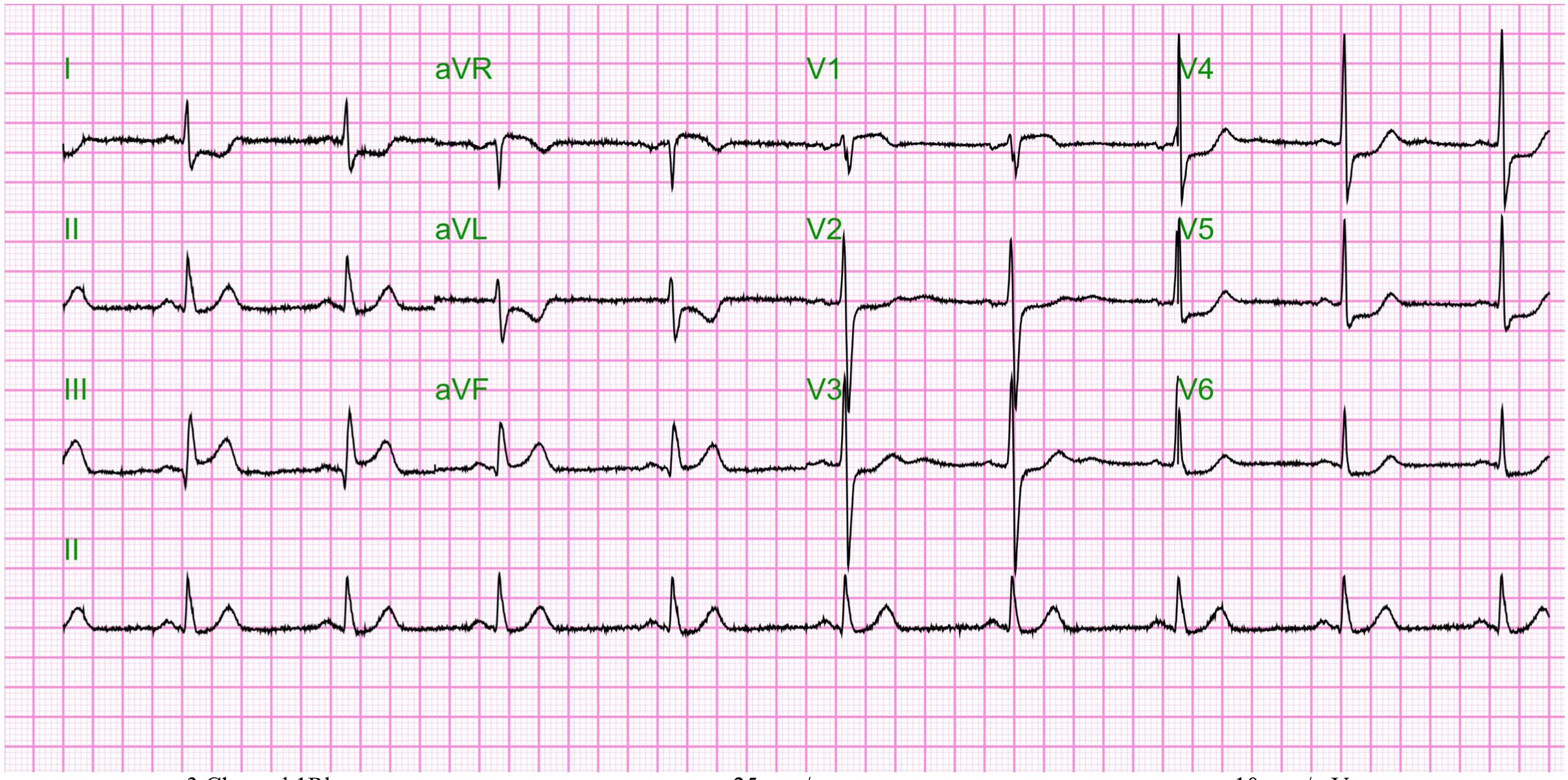
Relevant Test Results Prior to Catheterization
Relevant Catheterization Findings
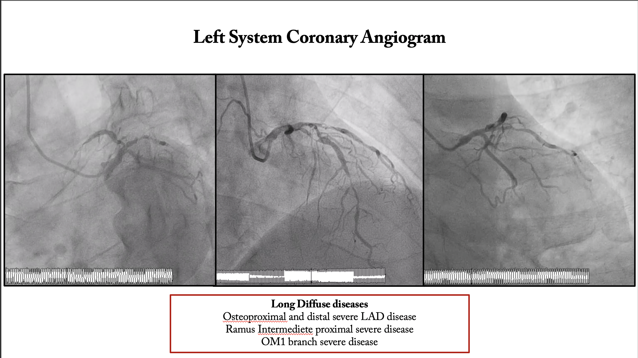
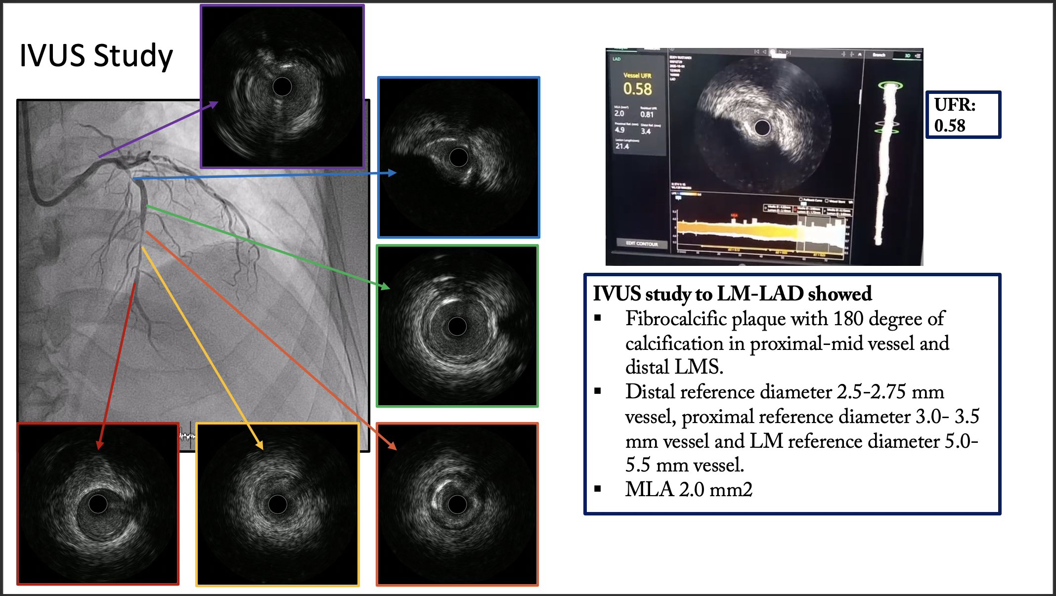
Coronary angiography revealed distal left main trifurcation disease, with significant stenosis involving the distal lm, ostial-to-proximal LAD, and proximal RI. The lesion was classified as modified medina 1-1-1-0. The syntax score was 30, consistent with a intermediete anatomical complexity, and the patient declined surgical revascularization (CABG). To preserve LCx and intermediate branch flow, PCI was performed using the simultaneous jailed balloon and jailed microcatheter technique.
 Spider view.mp4
RAO-cranial view.mp4
Interventional Management
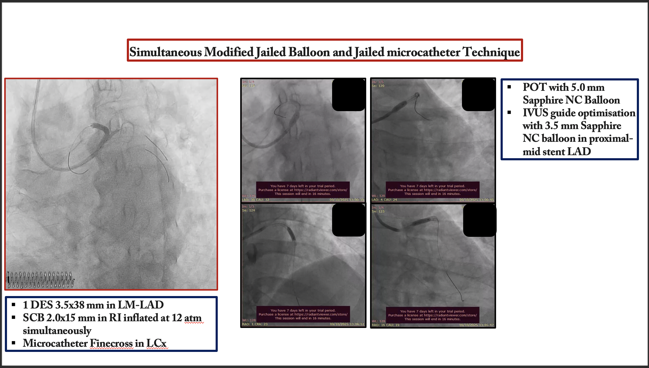
Procedural Step
An 7 Fr EBU 3.5 guiding catheter was engaged into the left coronary artery via the right radial artery. A SION guide wire was initially inserted into the LAD. To protect the two large side branches, a BMW guide wire and a runthrough with a Finecross microcatheter were introduced into the intermediate branch and LCX, respectively. According to the angiographic and intravascular ultrasound images, we decided to perform direct crossover stenting from the LMT to LAD using the simultaneous jailed balloon and jailed microcatheter technique. After lesions preparation, Ramus intermediete treated with 1 DEB 2.5x25 mm. A DES 3.5x38 was advanced into the LAD, and a semicompliant balloon 2.0 × 15 mm was advanced into the intermediate branch. Subsequently, a Finecross microcatheter was introduced into the LCX. The simultaneous jailed balloon and jailed Finecross technique was then performed. The side branch balloon in the intermediate branch was initially inflated at nominal pressure (12 atm). The main branch stent balloon in the LMT to LAD was then inflated at nominal pressure (11 atm), which simultaneously jailed the side branch semi-inflated balloon in the intermediate branch and the Finecross microcatheter in the LCX. Blood flow was preserved in both the intermediate branch and the LCX after main vessel stenting, and there were no signs of plaque or carina shift into the side branches. Distal LAD was treated with hybrid approach 1 DCB 2.5x35 mm.
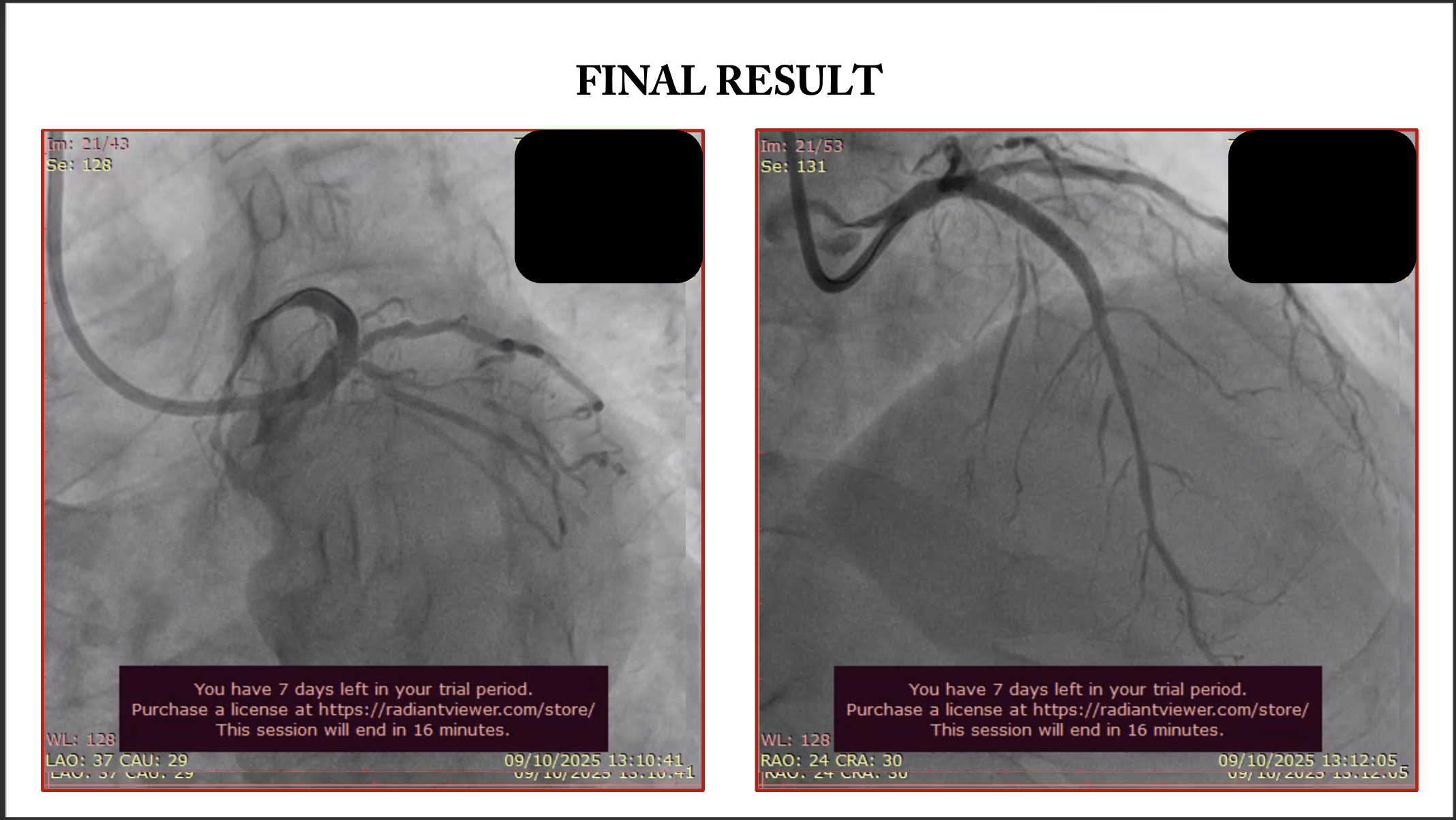
Final angiogram spider.mp4
Final Angiogram Cranial.mp4
Case Summary
The simultaneous jailed balloon and jailed microcatheter technique is a novel and effective double side branch protection technique for the treatment of left main trifurcation disease in selected patients. Further studies on this technique in larger populations are needed.