Lots of interesting abstracts and cases were submitted for TCTAP 2026. Below are the accepted ones after a thorough review by our official reviewers. Don’t miss the opportunity to expand your knowledge!
CASE20251111_002
Unexpected Stent Dislodgement During Percutaneous Coronary Intervention: Successful Retrieval and Lesson Learned
By Zheng Sheng Tan, De Zhi Law
Presenter
De Zhi Law
Authors
Zheng Sheng Tan1, De Zhi Law2
Affiliation
Hospital Sultan Idris Shah, Serdang, Malaysia1, Hospital Queen Elizabeth II, Malaysia2
View Study Report
CASE20251111_002
Coronary - Complication Management
Unexpected Stent Dislodgement During Percutaneous Coronary Intervention: Successful Retrieval and Lesson Learned
Zheng Sheng Tan1, De Zhi Law2
Hospital Sultan Idris Shah, Serdang, Malaysia1, Hospital Queen Elizabeth II, Malaysia2
Clinical Information
Relevant Clinical History and Physical Exam
50-year-oldgentleman with diabetes mellitus, hypertension, dyslipidaemia and ischemicheart disease presented to our centre for further management. He has past history of acute anterior myocardial infarction in June 2024 which was successfully thrombolysed and another episode of acute inferior myocardial infarction in June 2025 where he was thrombolysed again.

Relevant Test Results Prior to Catheterization
Echocardiogram: LVEF 58%, no regional wall motion abnormalities, no valve pathology
Relevant Catheterization Findings
Coronary angiogram:
LMS: normal
LAD: proximal 70% stenosis, mid 90% stenosis, distal 100% stenosisLCX: ostial chronic total occlusionRCA: ectatic, proximal 70% stenosis, distal 70% stenosis
LMS: normal
LAD: proximal 70% stenosis, mid 90% stenosis, distal 100% stenosisLCX: ostial chronic total occlusionRCA: ectatic, proximal 70% stenosis, distal 70% stenosis
Interventional Management
Procedural Step
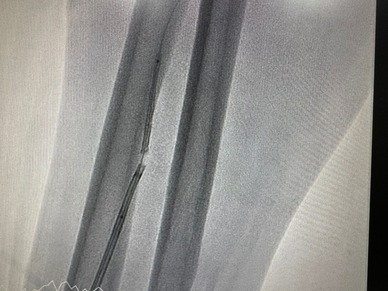
6Fr Guiding catheter JR 3.5 was used to engaged the rightcoronary artery and lesions was crossed with guidewire sion blue. Predilatationof lesions was performed sequentially with a 4.0 x 15mm non-compliant balloonthen 4.0 x 15mm scoring balloon, resulting in adequate lesion expansion andTIMI 3 flow. We proceeded to stent the mid to distal segment of RCA with 4.0 x36mm drug eluting stent. Subsequently attempted to stent the ostial to midsegment of RCA with 4.0 x 48mm durg eluting stent but while trying to readjustposition of stent, we were unable to withdraw the undeployed stent into theguiding catheter. Noted proximal stent edge crumpled at the mouth of guidingcatheter and stent balloon was retracted half into the guiding catheter. The whole system was pulled back into theradial artery where the stent balloon was pulled out of guiding catheter. Withthe stent still remained on the coronary guidewire, an amplatz gooseneck snarewas inserted. The crumpled stent was successfully snared out.
Case Summary
In our case, the most likely contributing factor wasnon-coaxiality between the guiding catheter and the right coronary arteryostium, which increased friction during device manipulation. This malalignmentmay have caused uneven force distribution when attempting to withdraw thestent, resulting in the stent detaching from the delivery balloon while theguidewire remained in place.