Lots of interesting abstracts and cases were submitted for TCTAP 2026. Below are the accepted ones after a thorough review by our official reviewers. Don’t miss the opportunity to expand your knowledge!
CASE20251110_007
Two-Dimensional/Three-Dimensional Registration for Fusion Imaging in Renal Endovascular Intervention
By Erwin Mulia, Suko Adiarto
Presenter
Erwin Mulia
Authors
Erwin Mulia1, Suko Adiarto1
Affiliation
Harapan Kita Hospital, Indonesia1
View Study Report
CASE20251110_007
Endovascular - Other Endovascular Interventions
Two-Dimensional/Three-Dimensional Registration for Fusion Imaging in Renal Endovascular Intervention
Erwin Mulia1, Suko Adiarto1
Harapan Kita Hospital, Indonesia1
Clinical Information
Relevant Clinical History and Physical Exam
Fourteen year old boy was consulted to our Vascular Medicine Department because of the uncontrolled hypertension. He was treated with amlodipine and clonidine from Pediatric Department. He had history of ventricular septal defect which was closed percutaneously. Despite having escalated anti hypertension medication, the blood pressure seemed difficult to control. Blood pressure during admission was 174/87 mmHg and heart rate was 73 beat per minute. Body mass index was 16.2 kg/ m2.
Relevant Test Results Prior to Catheterization
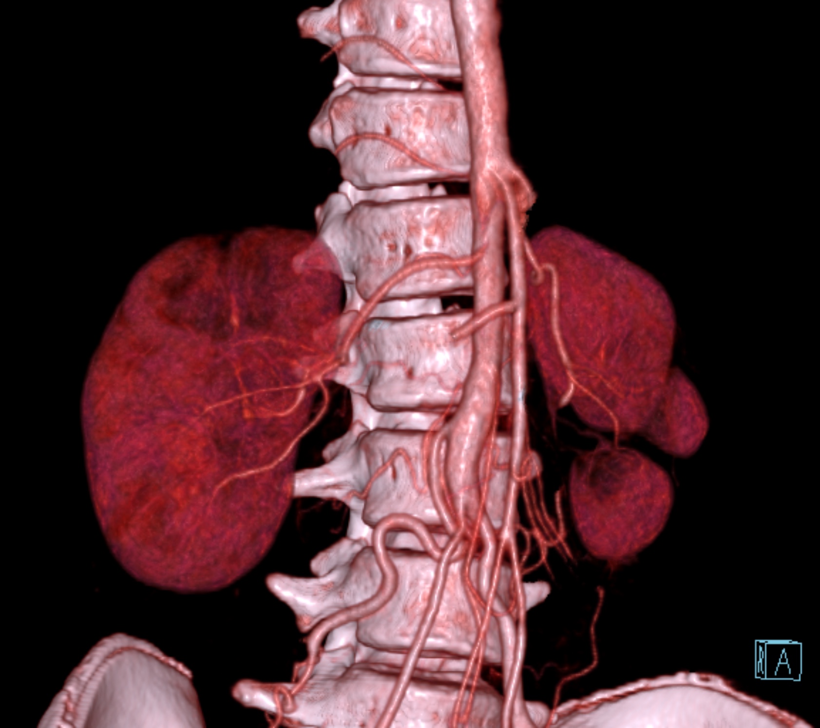
Echocardiogram showed preserved left ventricular ejection fraction with mild aortic regurgitation without residual VSD. Laboratory results showed normal renal function with estimated glomerular filtration rate of more than 75. Computed tomography angiography showed significant stenosis at the ostium of right renal artery.
 movie_20250806_15-24-14.mp4
movie_20250806_15-08-09.mp4
Relevant Catheterization Findings
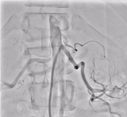
Six french femoral arterial sheath was placed under ultrasound guided. Renal angiogram using 6 Fr Renal Double Curve catheter showed normal left renal artery and subtotal occlusion near at the ostium of right renal artery.
imitation VE.mp4
Frontal.dcm.mov
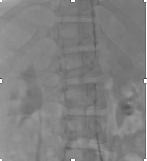
Interventional Management
Procedural Step
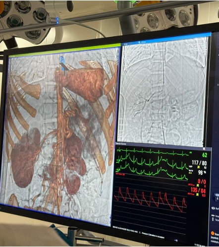
Despite having an overlay from three dimensional three dimensional fusion imaging, it was difficult to wire the right renal artery. Five French right brachial arterial sheath was introduced under ultrasound guided. Polymer jacket coronary guide wire was used under two dimensional three dimensional fusion imaging. Wiring was finally succeeded with the support of JR 3.5/ 5 Fr guiding catheter from the brachial access. Lesion was predilated using 2.0 x 15 mm and 4.0 x 15 mm semi compliant coronary balloons. Drug eluting stent of 4.0 x 15 mm was implanted and post dilated using 4.5 x 12 mm non compliant coronary balloon. The procedure was uneventful. Fluoroscopy time was 53.16 minutes, contrast volume was 130 cc, radiation dose was 83.88 mGy.
IMI4.mp4
IMI6.mp4
IMI11.mp4
Case Summary
We have presented case of uncontrolled hypertension in 14 year old boy due to right renal artery stenosis. The renal endovascular intervention was performed using two dimensional three dimensional fusion imaging technique. Despite the high and tight origin of the right renal artery, the procedure was completed successfully. In 1.5 months follow up, the blood pressure was easily controlled and the medication could be deescalated. Two dimensional three dimensional fusion imaging is one technique needed when compared with three dimensional three dimensional fusion imaging to reduce the radiation dose, contrast usage, especially in difficult procedural scenario like stated in this case.