Lots of interesting abstracts and cases were submitted for TCTAP 2026. Below are the accepted ones after a thorough review by our official reviewers. Don’t miss the opportunity to expand your knowledge!
CASE20251107_031
The Suture of Life: Type V Coronary Perforation Management With Chromic Catgut Embolization
By Robin Hendra Wibowo
Presenter
Robin Hendra Wibowo
Authors
Robin Hendra Wibowo1
Affiliation
Pasundan University, Indonesia1
View Study Report
CASE20251107_031
Coronary - Complication Management
The Suture of Life: Type V Coronary Perforation Management With Chromic Catgut Embolization
Robin Hendra Wibowo1
Pasundan University, Indonesia1
Clinical Information
Relevant Clinical History and Physical Exam
A 56-year-old female with history of hypertension and dyslipimdeia presented to the emergency department complaining of chest pain and diaphoresis. Vital signs at admission and physical examination were unremarkable.
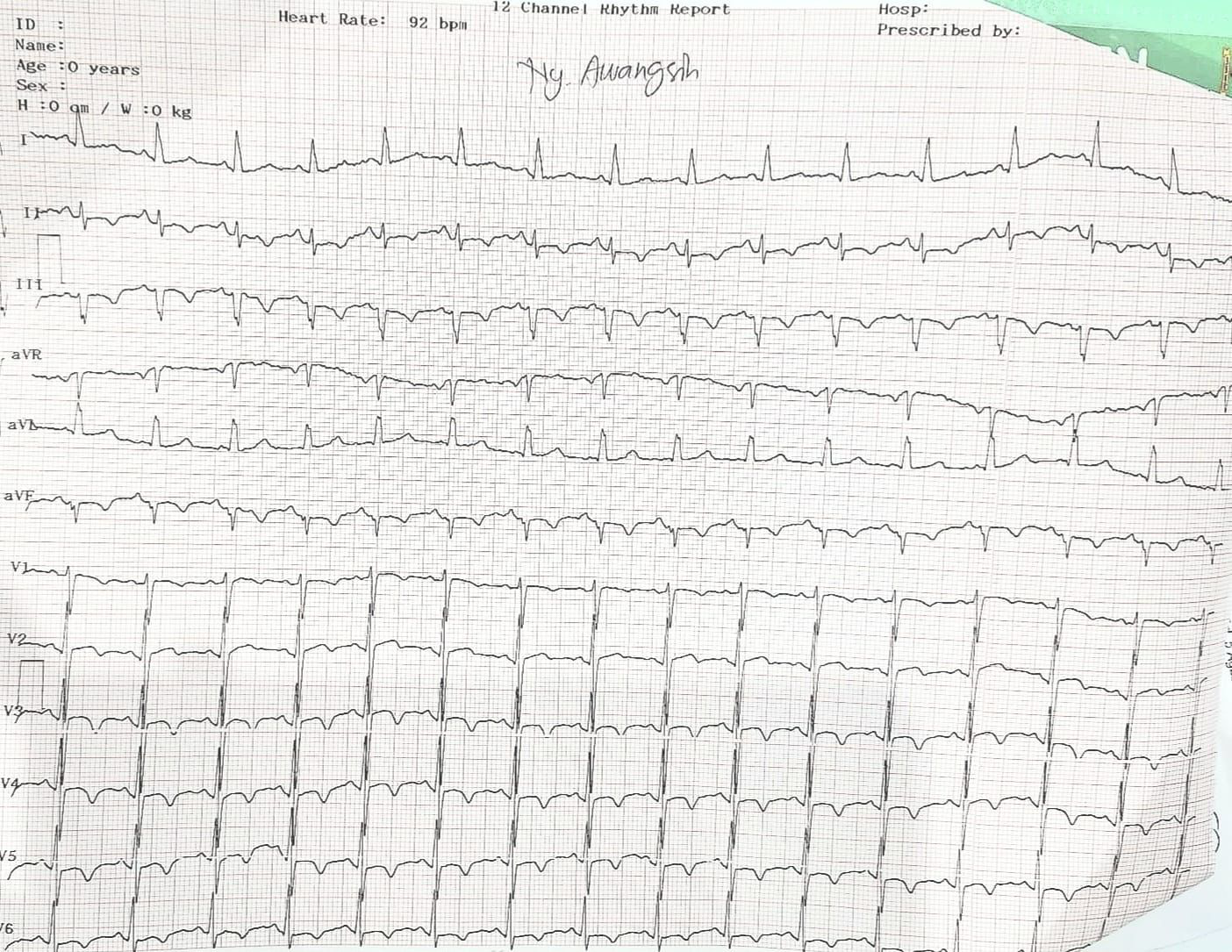
Relevant Test Results Prior to Catheterization
Her Electrocardiogram (ECG) showed sinus rhythm, ST segment depression in infero-anterior segments with elevated troponin I level (78ng/L). The diagnosis of NSTEMI infero-anterior was established and referred to the cathlab for urgent PCI.

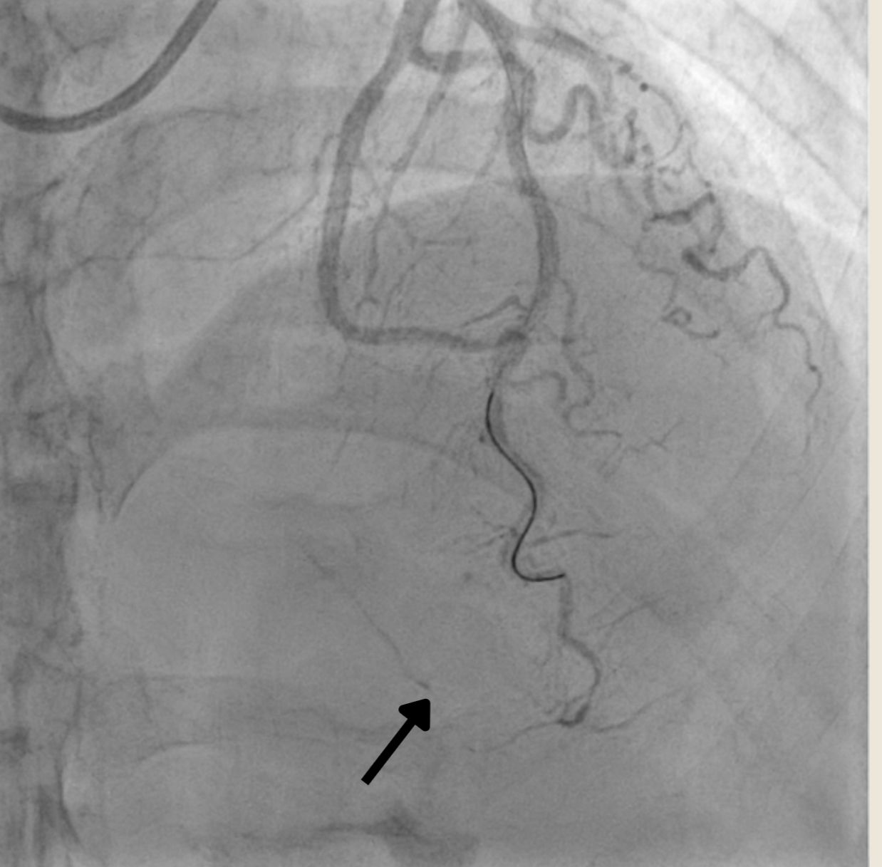
Relevant Catheterization Findings
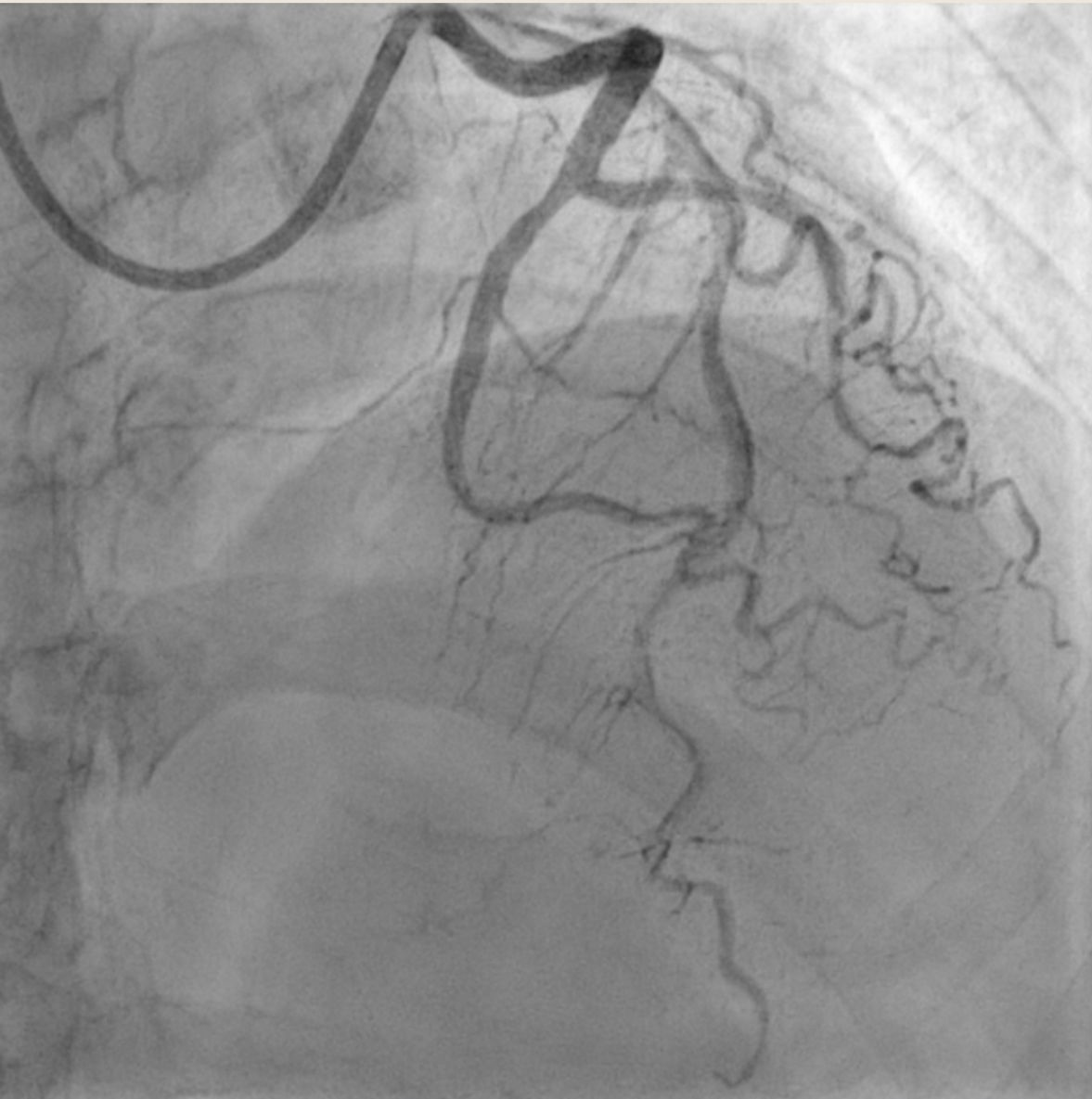
Angiography revealed single vessel disease. Left anterior descending artery (LAD) showed mild stenosis of proximal-mid part and total occlusion of the distal part Left circumflex (LCx) artery and The right coronary artery (RCA) showed minimally diseased.
Interventional Management
Procedural Step
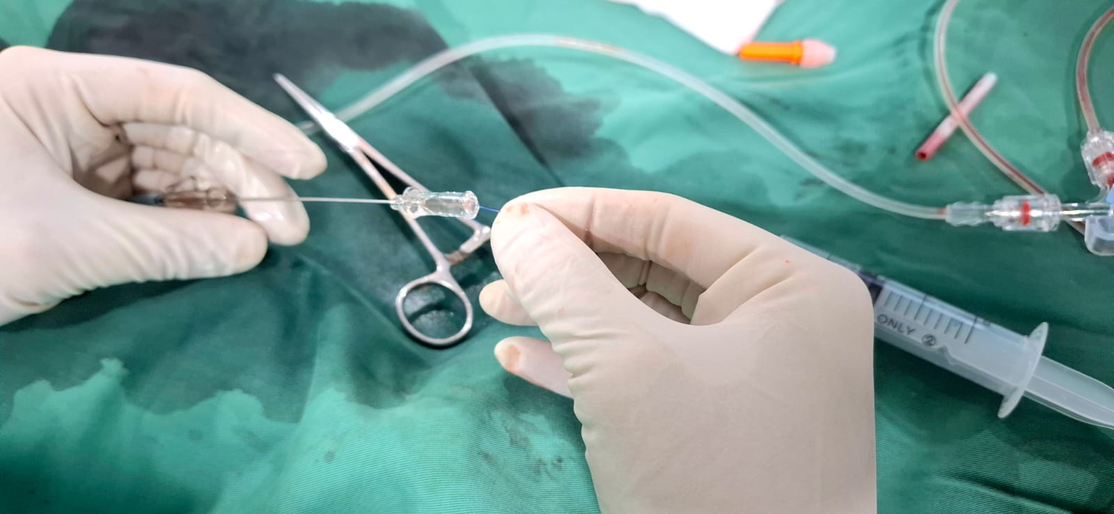
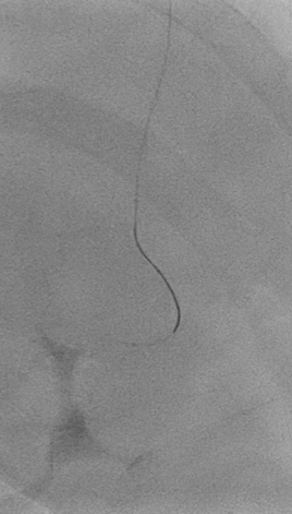
PCI was conducted using guide catheter EBU3.5/6F, A 0.014-inch floppy guidewire and was placed in distal LAD. After sliding the distal lesion with semicompliant 1.5 x 15mm, we encountered distal perforation type V with Ellis type III. Multiple prolonged ballon inflation was done using semicompliant 2.0 x 15mm at 4 atm but the perforation jet persisted. We decided to do microcatheter-assisted absorbable suture embolization technique with chromic catgut 3.0. The perforation was successfully managed. Echocardiography evaluation revealed Concentric Left Ventricular Hyperthrophy with normal LVEF without signs of pericardial effusion. Patient was dismissed from hospitalization without further complication.
Case Summary
We reported a 56-year-old female with NSTEMI and PCI was done with Type V coronary complication and successfully sealed with Chromic Catgut suture embolization. the use of chromic gut suture for sealing Type V coronary perforations appears to be a feasible and safe bailout option when conventional methods are unsuccessful or not applicable. The technique provides targeted, distal microvascular embolization. However, its efficacy and long-term safety remain unproven. Large-scale registries or randomized trials are warranted but are challenging due to the rarity of the event.