E-Case

Lots of interesting abstracts and cases were submitted for TCTAP 2026. Below are the accepted ones after a thorough review by our official reviewers. Don¡¯t miss the opportunity to expand your knowledge!

CASE20251107_003

Take a Detour in Critical LM Disease and Long Calcified LAD

By Yen-Lien Chou

Presenter

Yen-Lien Chou

Authors

Yen-Lien Chou1

Affiliation

Tri-Service General Hospital, Taiwan1
View Study Report
CASE20251107_003
Coronary - Complex PCI - Calcified Lesion

Take a Detour in Critical LM Disease and Long Calcified LAD

Yen-Lien Chou1

Tri-Service General Hospital, Taiwan1

Clinical Information

Relevant Clinical History and Physical Exam

64-year-old man, ex-smoker, Hx ofspontaneous ICH, HTN, and BPHFather died due to AMI and Mother with CVA and HTNIntermittent chest pain 2months ago and frequent taking sublingual NTGThe symptoms attack more frequently and accompanyingwith cold sweating

Relevant Test Results Prior to Catheterization

HDLC 44;LDLC 101;GLU 93; HBA1C5.5; CREA 0.9;AST 19;ALT 13Echocardiography showed Mid-reducedLVSF with EF: 40%, Mild MR, Hypokinesiaof anterior wall, and PAP: 27mmHg

Relevant Catheterization Findings

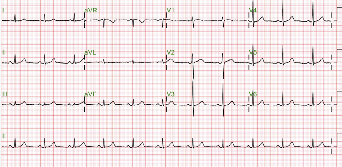
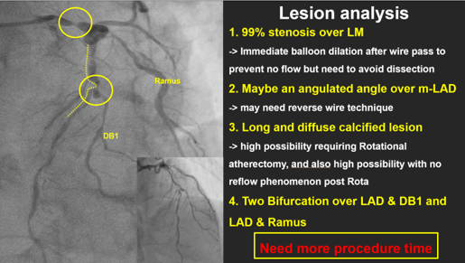
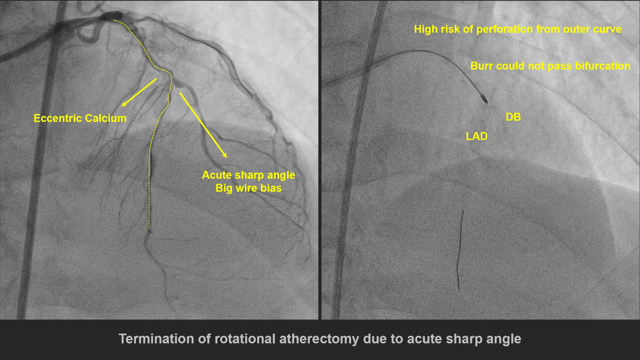
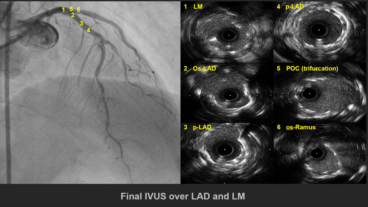
LM: D/3:discrete 99% stenosis, Medina (1,0,0) with LAD and LCxLAD: heavycalcificationP/3 to M/3: Diffuse60-95% stenosis, Medina (1,1,0) with DB1 (aneurysm formation over ostium)D/3: Diffuse 60-70% stenosisP/3 to M/3 of DB1: Tubular 70-80%stenosisLCx: Patentand non-dominant vesselRamus Intermedius : Ostium to M/3: Diffuse 70-95% stenosisRCA: calcificationD/3: Diffuse 70-80% stenosis P/3 of PDA: Discrete 60-70% stenosisGiving collateral flow to septalperforator


ØÚô÷1.mp4
ØÚô÷2.mp4
ØÚô÷3.mp4

Interventional Management

Procedural Step

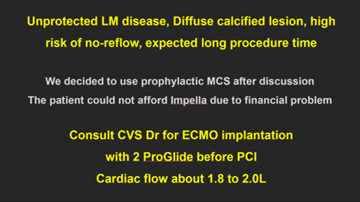
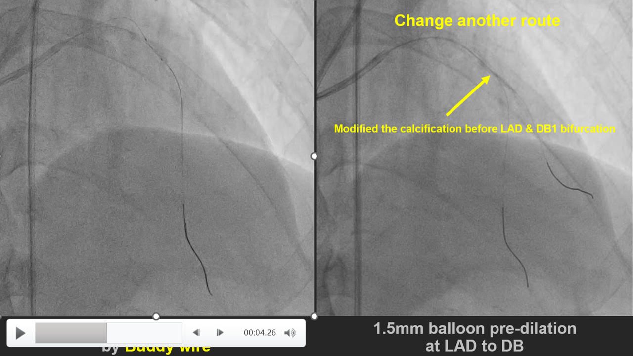
1. V-A mode ECMO was used for CHIP PCI2. 1.5mm balloon was advanced to m-LAD3. 2.0mm balloon failed to cross the heavycalcified lesion at m-LAD4. By Extra support rota wire, 1.25mm Burradvanced to the lesion in a slow and steady manner (163300-182000 rpm). p-LADlesion was polished, however, Burr could not pass through m-LAD tortuous andheavy calcified bifurcation lesion. To avoid high risk of perforation, weterminated the Rotational Atherectomy5. 1.5mm balloon was advanced to m-LADlesion6. 2.0 balloon failed to deliver to the M/3LAD tortuous and heavy calcified bifurcation lesion, even under 5.5 FrGuideLiner or Buddy wire technique7. 2.0mm balloon was advanced to m-LAD throughDB wire, inflated at pressure 22-24 atm8. 2.0mm Cutting balloon was advanced tothe lesion through DB wire, inflated at pressure 6-18 atm9. 2.0mm balloon was advanced to the lesionthrough DB wire10. A 2.0 balloon successfully advanced to mto d-LAD11. 2.0mm Cutting balloon was advanced tothe lesion through LAD wire, inflated at pressure 8-18 atm12. 2.5mm balloon was advanced to m-LADlesion13. 2.0x30mm Onyx stent failed to adavnceto the lesion, even under GuideLiner 6Fr14. Finally 2.0x30mm Onyx stentsuccessfully advanced to the proper segment at m-LAD under Wiggle wire support15. 2.5x28mm Xience Sierra stent wasdeployed at p-Ramus16. 2.75x30mm Onyx stent was deployed at LMto m-LAD17. Final angiography showed TIMI 3 distalflow & No extravasation


ØÚô÷4.mp4
ØÚô÷5.mp4
ØÚô÷6.mp4

Case Summary

*. Critical LM disease with longcalcified lesion may need more procedure time, prophylactic MCS should beconsidered*. Rotational atherectomy in Angulatedand calcified lesion should be carful with complication, half-way rotationalatherecmoy should be considered*. Angulated and calcified lesion, usingCutting and NC balloon could be an another choice to modified lesion. If a bigside branch (diameter > 2.5mm) after angulated and calcified lesion may be anotherway to modified plque.*. Provisional stenting technique inbifurcation should be a first choice to make procedure simple and saveprocedure time