Lots of interesting abstracts and cases were submitted for TCTAP 2026. Below are the accepted ones after a thorough review by our official reviewers. Don’t miss the opportunity to expand your knowledge!
CASE20251106_025
First CT-Guided Percutaneous Coronary Intervention in Asia: Participation in the P4 Study
By Hirofumi Ohashi, Atomu Tajima, Koshiro Sakai, Takuya Mizukami, Hirohiko Ando, Carlos Collet, Tetsuya Amano
Presenter
Hirofumi Ohashi
Authors
Hirofumi Ohashi1, Atomu Tajima1, Koshiro Sakai2, Takuya Mizukami2, Hirohiko Ando1, Carlos Collet3, Tetsuya Amano1
Affiliation
Aichi Medical University, Japan1, Showa Medical University, Japan2, The Cardiovascular Research Foundation, USA3
View Study Report
CASE20251106_025
Coronary - Imaging & Physiology - Non-Invasive Imaging (CTA, MRI, Echo, etc)
First CT-Guided Percutaneous Coronary Intervention in Asia: Participation in the P4 Study
Hirofumi Ohashi1, Atomu Tajima1, Koshiro Sakai2, Takuya Mizukami2, Hirohiko Ando1, Carlos Collet3, Tetsuya Amano1
Aichi Medical University, Japan1, Showa Medical University, Japan2, The Cardiovascular Research Foundation, USA3
Clinical Information
Relevant Clinical History and Physical Exam
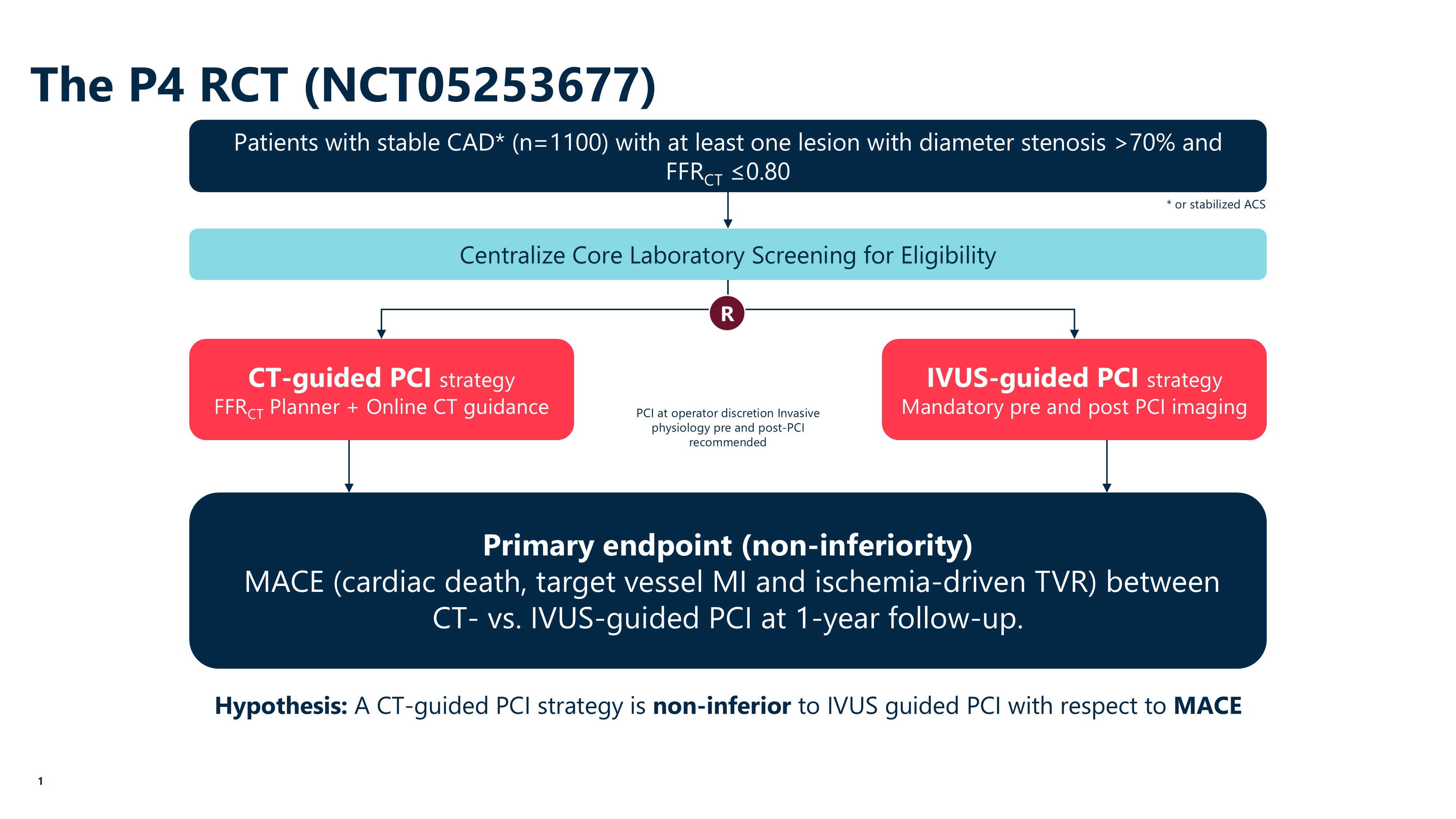
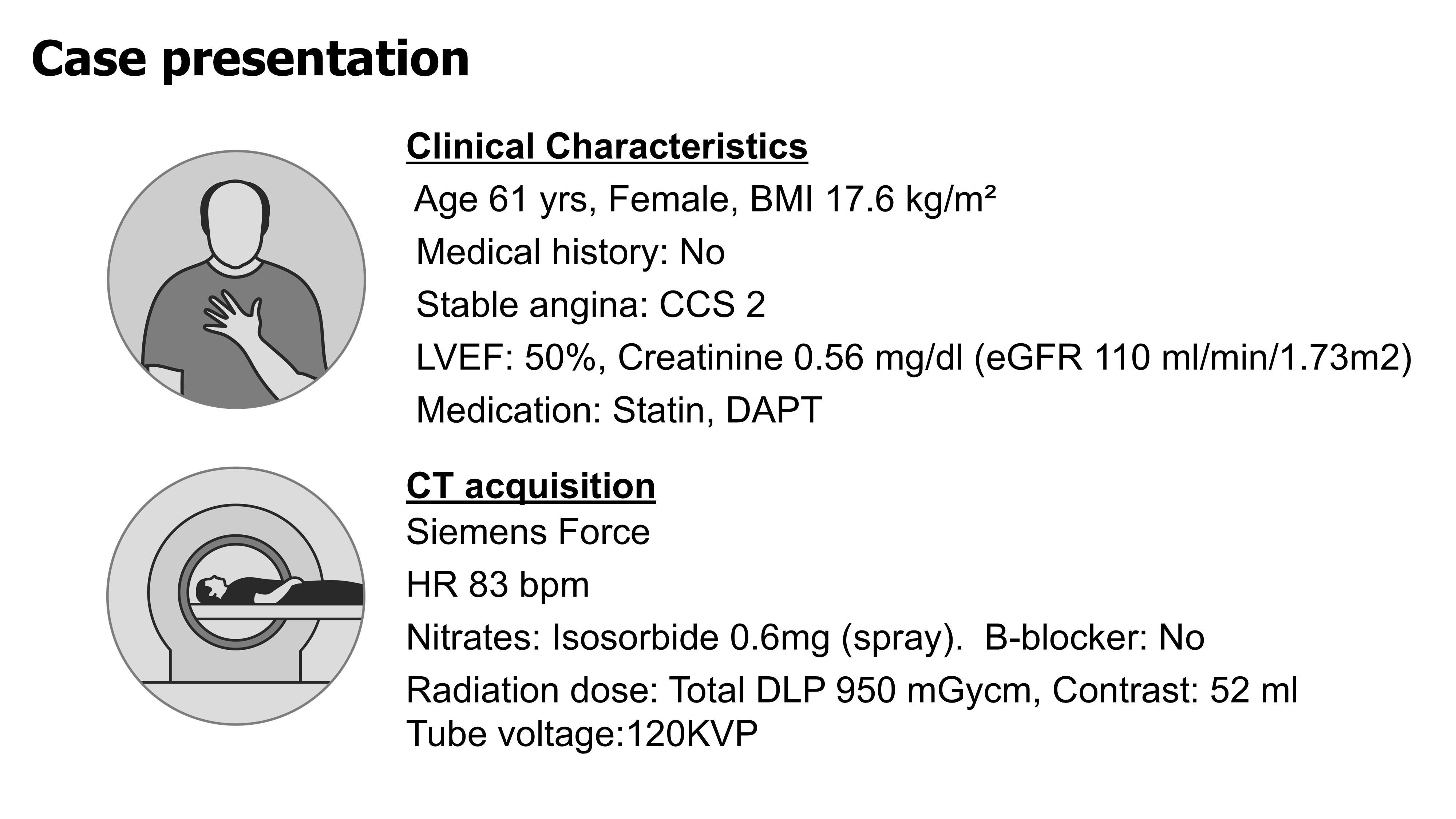
A 58-year-old woman with hyperlipidemia and stable angina was enrolled in the P4 study, a global randomized trial comparing CT-guided and IVUS-guided PCI. She had no diabetes or renal dysfunction. Physical examination and echocardiography were unremarkable. Baseline medications included aspirin and statin.
Relevant Test Results Prior to Catheterization
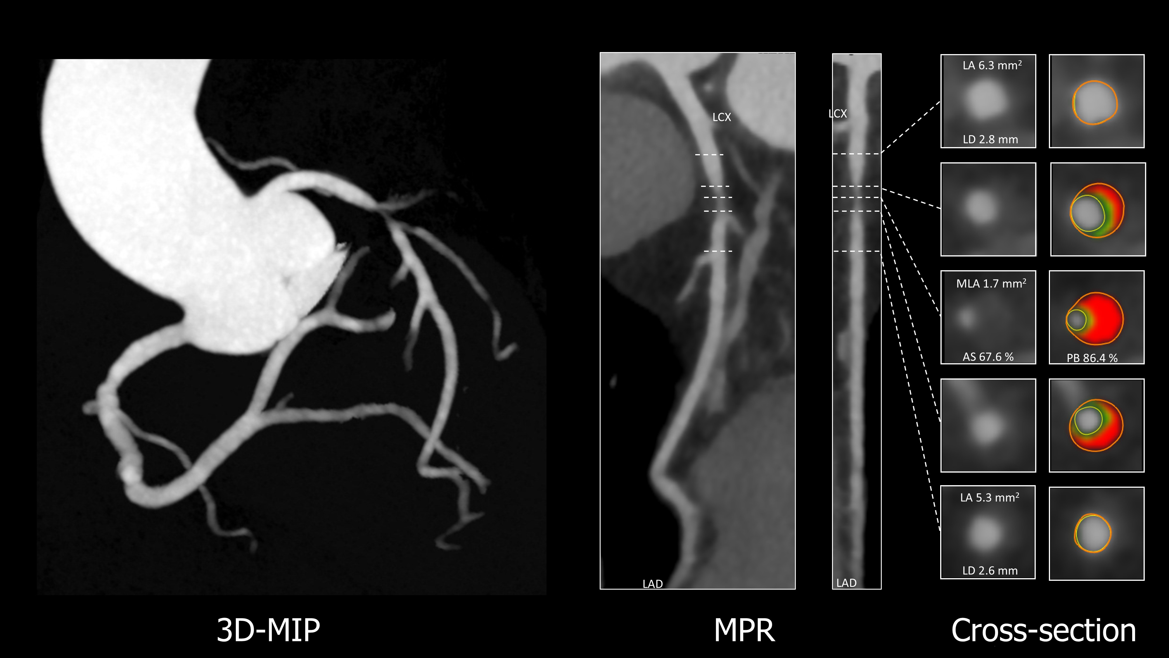
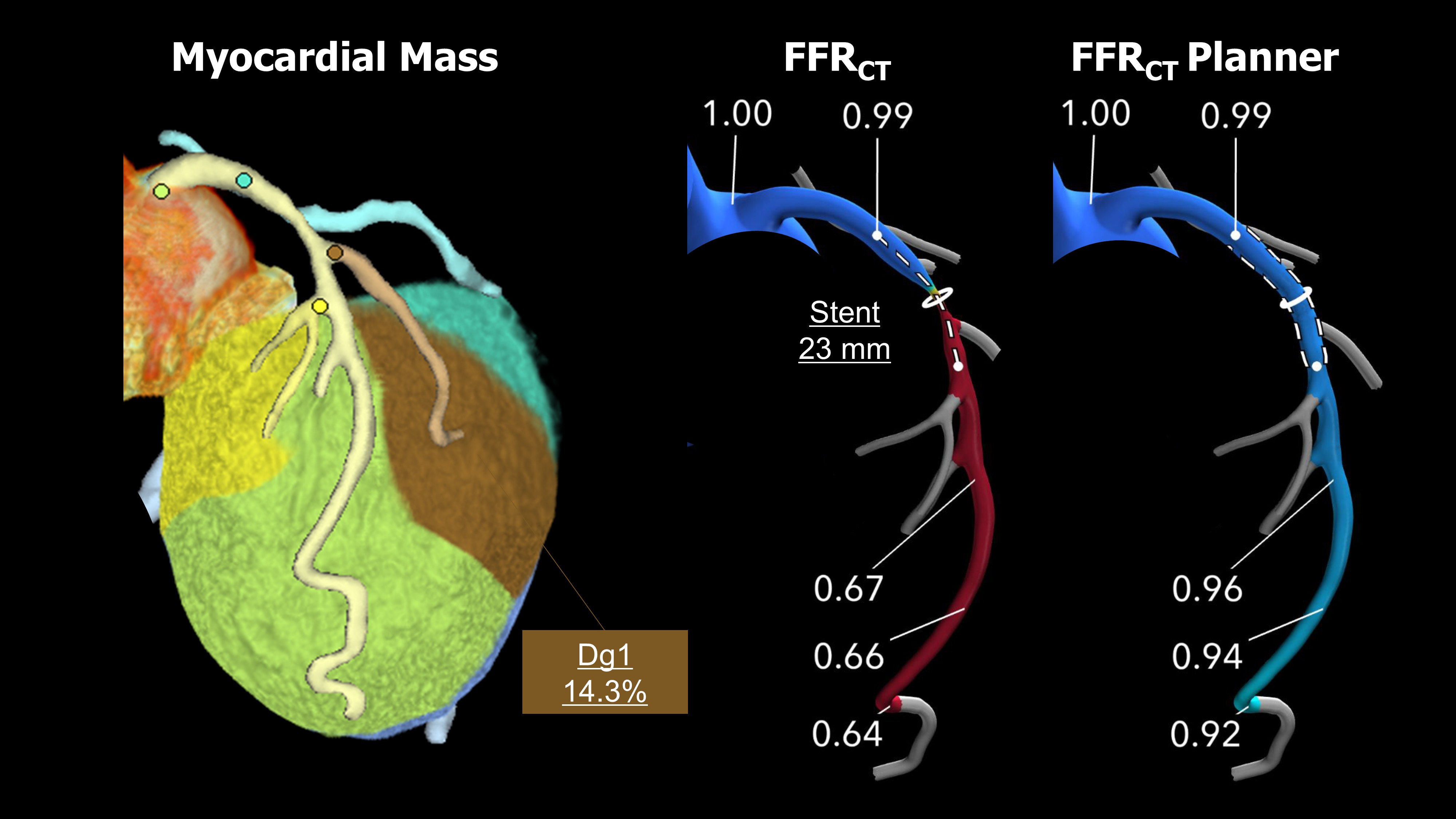
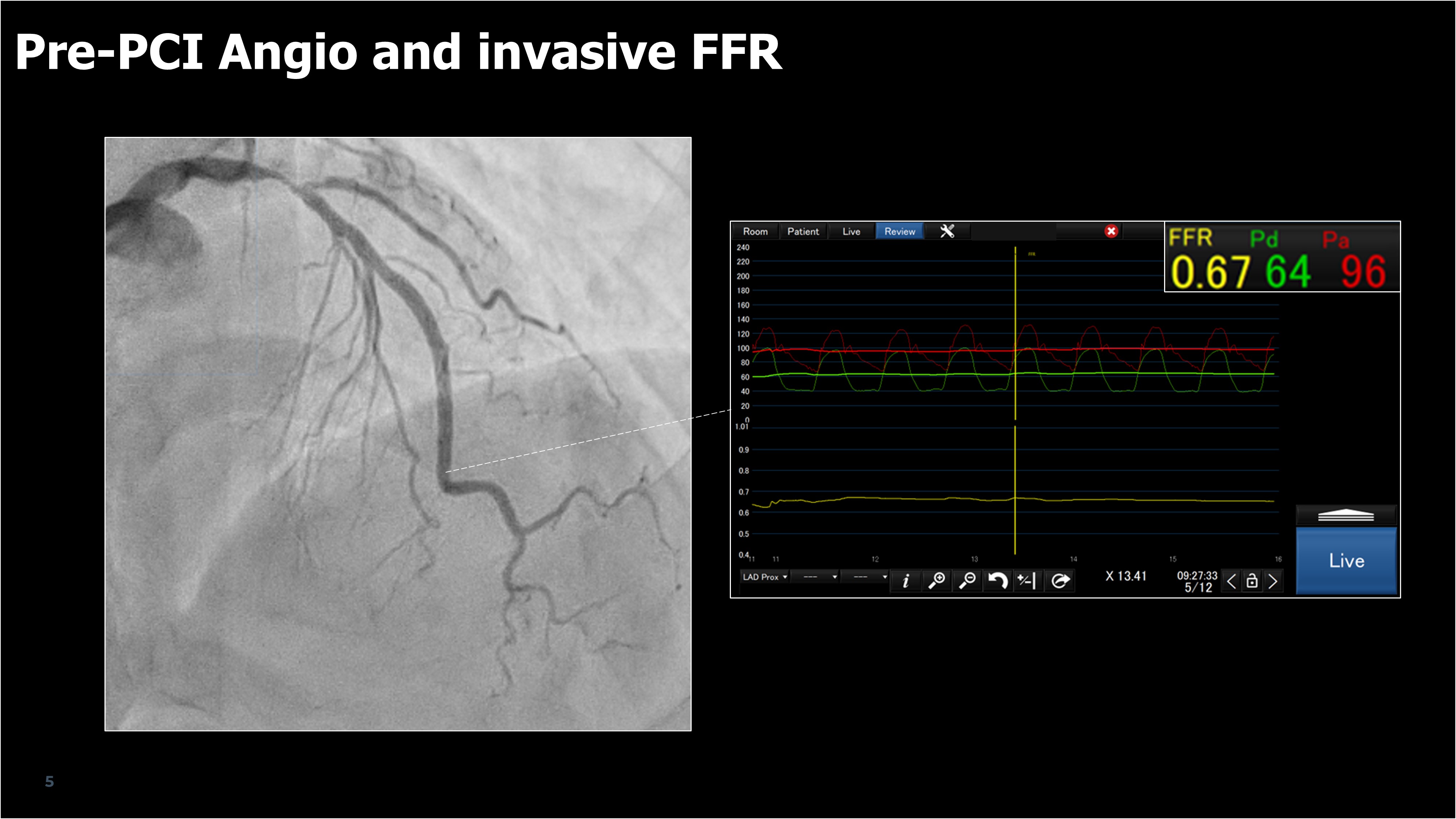
Coronary CT angiography (CCTA) demonstrated significant stenosis in the proximal left anterior descending artery (LAD). CT-derived fractional flow reserve (FFRCT) showed ischemia (0.68). CT analysis guided stent sizing with 23mm. The lesion was predominantly lipid-rich without calcification, requiring no modification. Virtual PCI using the FFRCT Planner predicted post-PCI FFRCT of 0.92.
Relevant Catheterization Findings
Coronary angiography confirmed severe proximal LAD stenosis consistent with the CCTA findings. Invasive FFR before PCI was 0.67.
Interventional Management
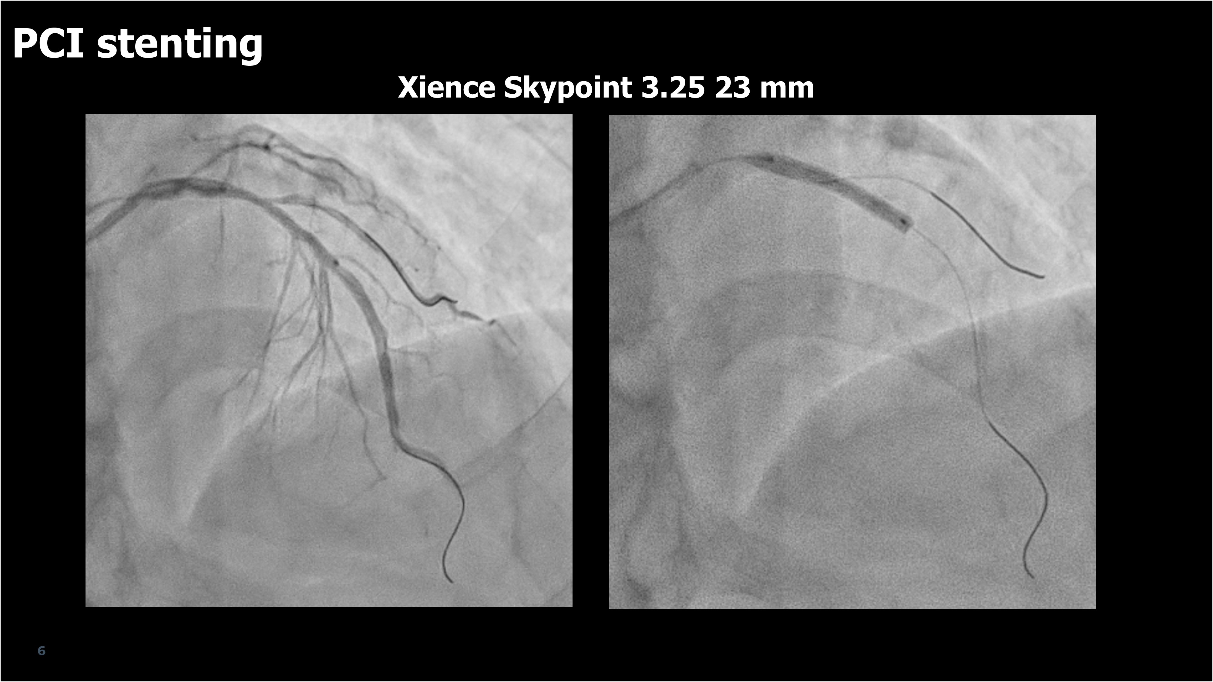
Procedural Step
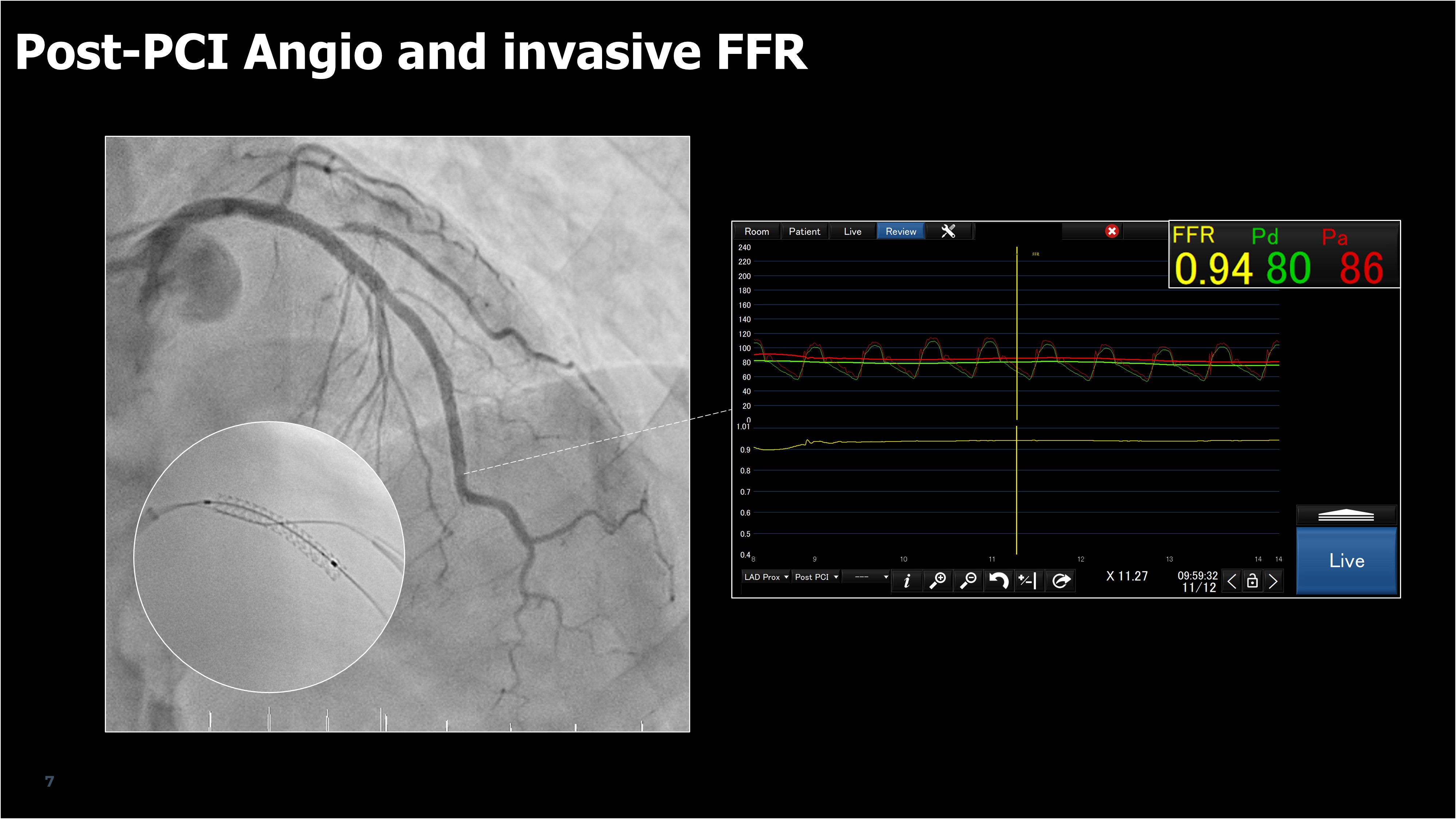
Following the CT-guided plan, PCI was performed without intravascular imaging, in accordance with the P4 study protocol.A 3.0 × 23 mm drug-eluting stent (Xience Skypoint, Abbott Vascular, USA) was deployed in the proximal LAD, with wire protection for the diagonal branch supplying 14% of myocardial mass. Post-dilatation achieved optimal expansion confirmed by stent enhancement. The final angiogram showed excellent result with preserved side branch flow.Invasive FFR improved from 0.67 to 0.94, closely matching the predicted FFRCT value (0.92). The procedure was uneventful, and no complications occurred during 30-day follow-up.
Case Summary
CT-guided PCI enabled precise pre-procedural planning, including lesion assessment, stent sizing, and branch evaluation. The results showed excellent agreement between predicted and measured FFR, demonstrating both the feasibility and accuracy of CT-based planning. This first Asian case within the P4 study highlights the potential of CT-guided PCI as a safe, non-invasive, and standardized alternative to intravascular imaging.