Lots of interesting abstracts and cases were submitted for TCTAP 2026. Below are the accepted ones after a thorough review by our official reviewers. Don’t miss the opportunity to expand your knowledge!
CASE20251104_013
A Case of Acute Coronary Syndrome in Pregnancy
By Jiho Han, Margaret McEntegart
Presenter
Jiho Han
Authors
Jiho Han1, Margaret McEntegart1
Affiliation
Columbia University, USA1
View Study Report
CASE20251104_013
Coronary - Complex PCI - Left Main
A Case of Acute Coronary Syndrome in Pregnancy
Jiho Han1, Margaret McEntegart1
Columbia University, USA1
Clinical Information
Relevant Clinical History and Physical Exam
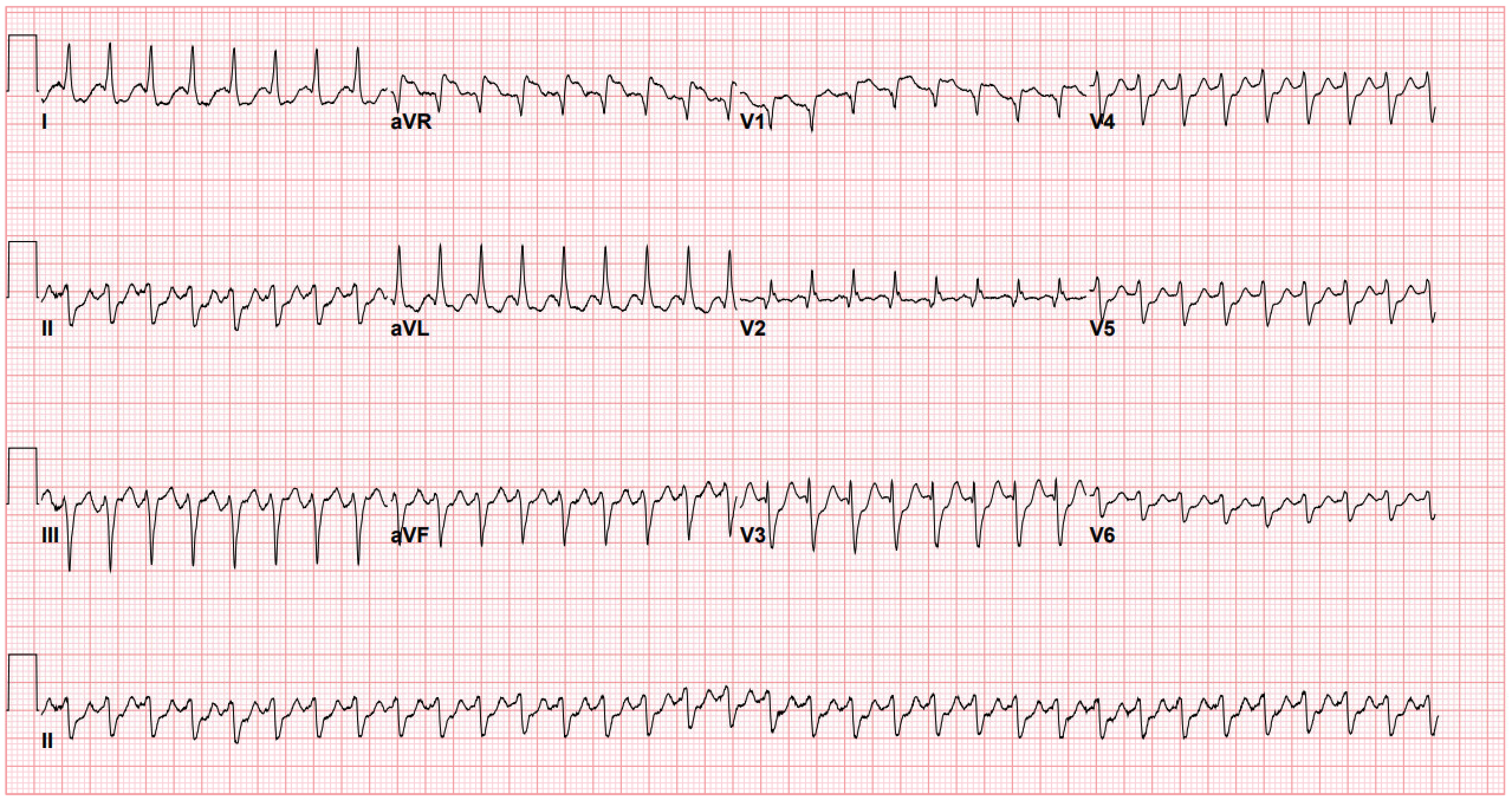
36 year old female G3P1102 with past medical history of non-Hodgkin's lymphoma (s/p chemotherapy x2, radiation therapy x40 sessions, experimental immunotherapies x2, BMT- in remission since 2016), MGUS, active pregnancy (20 weeks), pre-eclampsia who presented to an outside institution with palpitations, found to have SVT, ST elevations, and rising high-sensitivity troponin and transferred to CCU at our institution for further management
Relevant Test Results Prior to Catheterization
Echocardiogram showed mildly reduced left ventricular ejection fraction (45-50%) with segmental wall motion abnormalities with moderate hypokinesis of the mid to apical anteroseptal, mid to apical anterior, and apical myocardium without significant valvular disease. OB ultrasound showed intra-uterine pregnancy, normal fetal growth. Labs notable for high-sensitivity troponin peak at 25865 ng/L, creatinine 0.9 mg/dL, hemoglobin, 9.7 g/dL (baseline).
Relevant Catheterization Findings
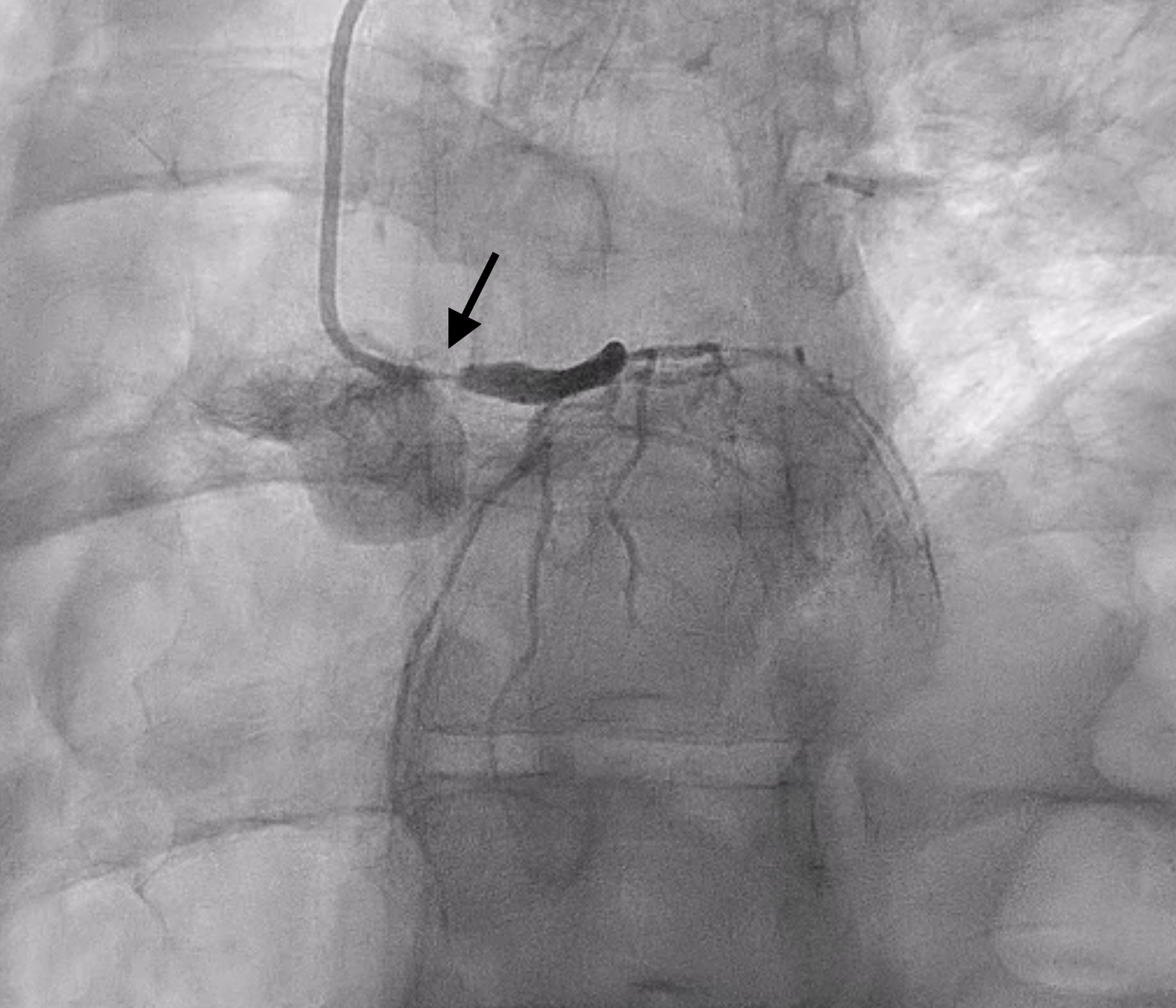
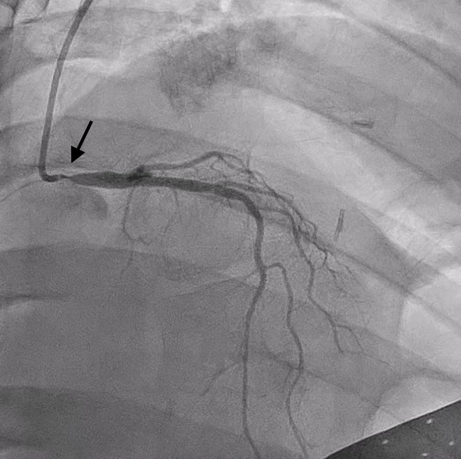
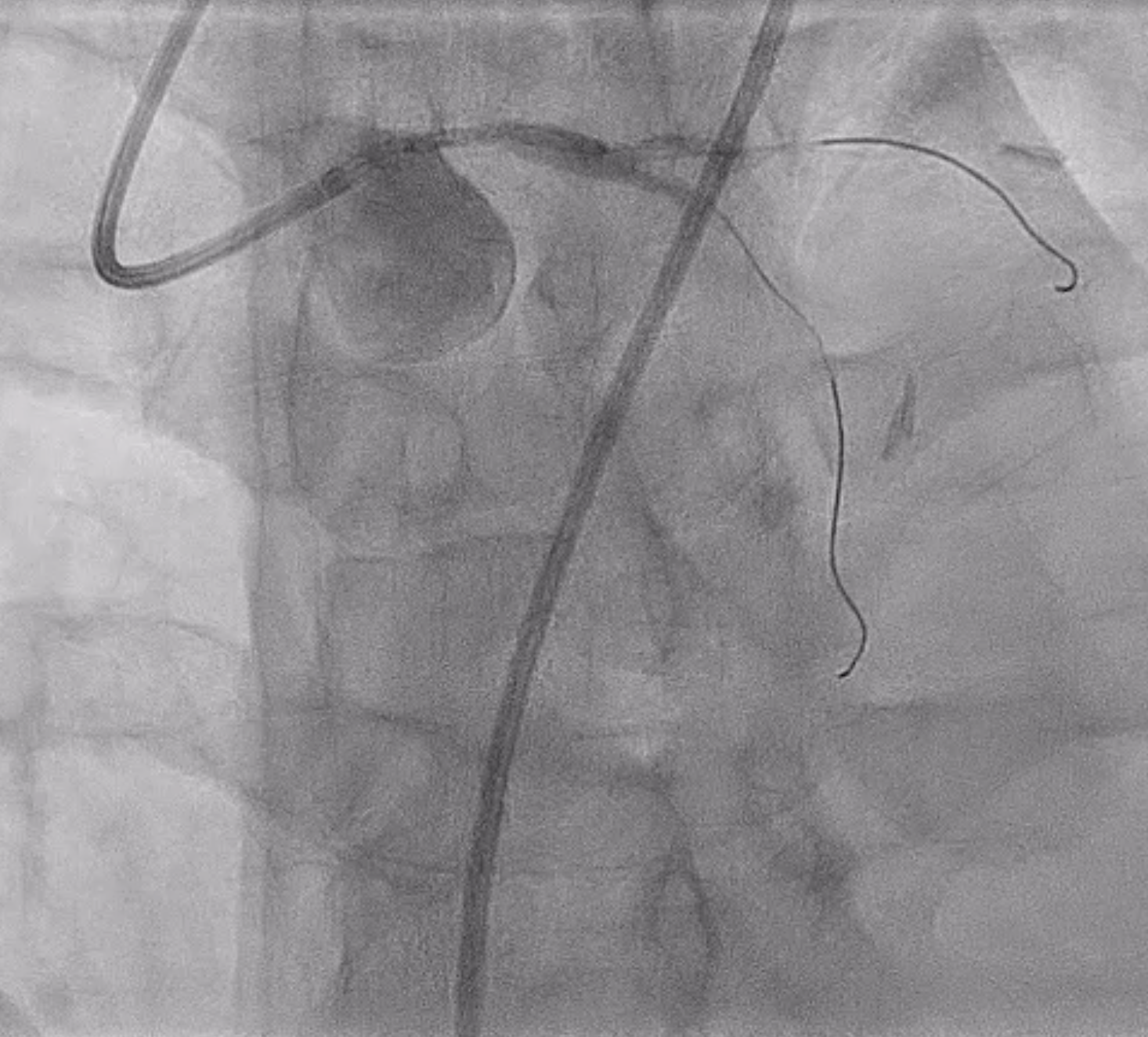
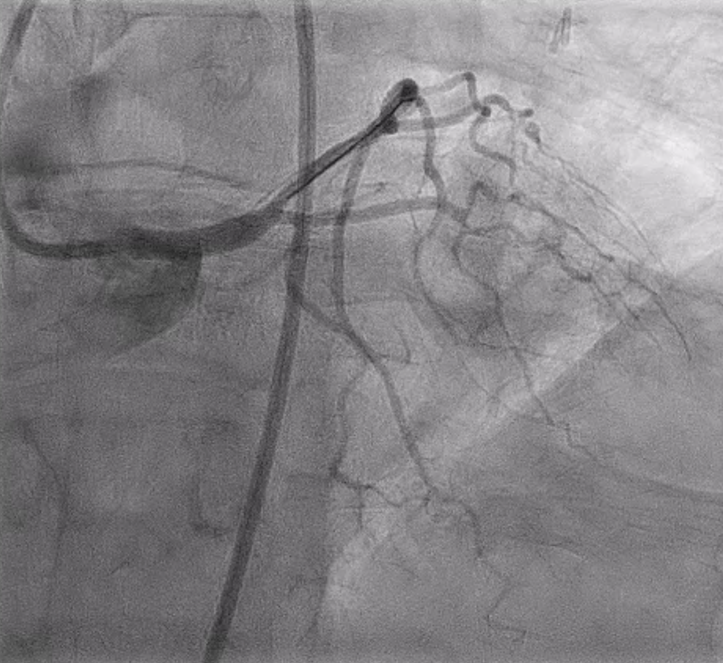
Left main has ostial to mid lesion with 90-95% stenosis. Left anterior descending and left circumflex and right coronary arteries have luminal irregularities without angiographic evidence of obstructive disease. Extreme angles were avoided during diagnostic angiogram in setting of her active pregnancy.
 Diagnostic Angiogram LAO20CRA15.mp4
Diagnostic Angiogram LAO21.mp4
Diagnostic Angiogram RAO20CRA20.mp4
Interventional Management
Procedural Step
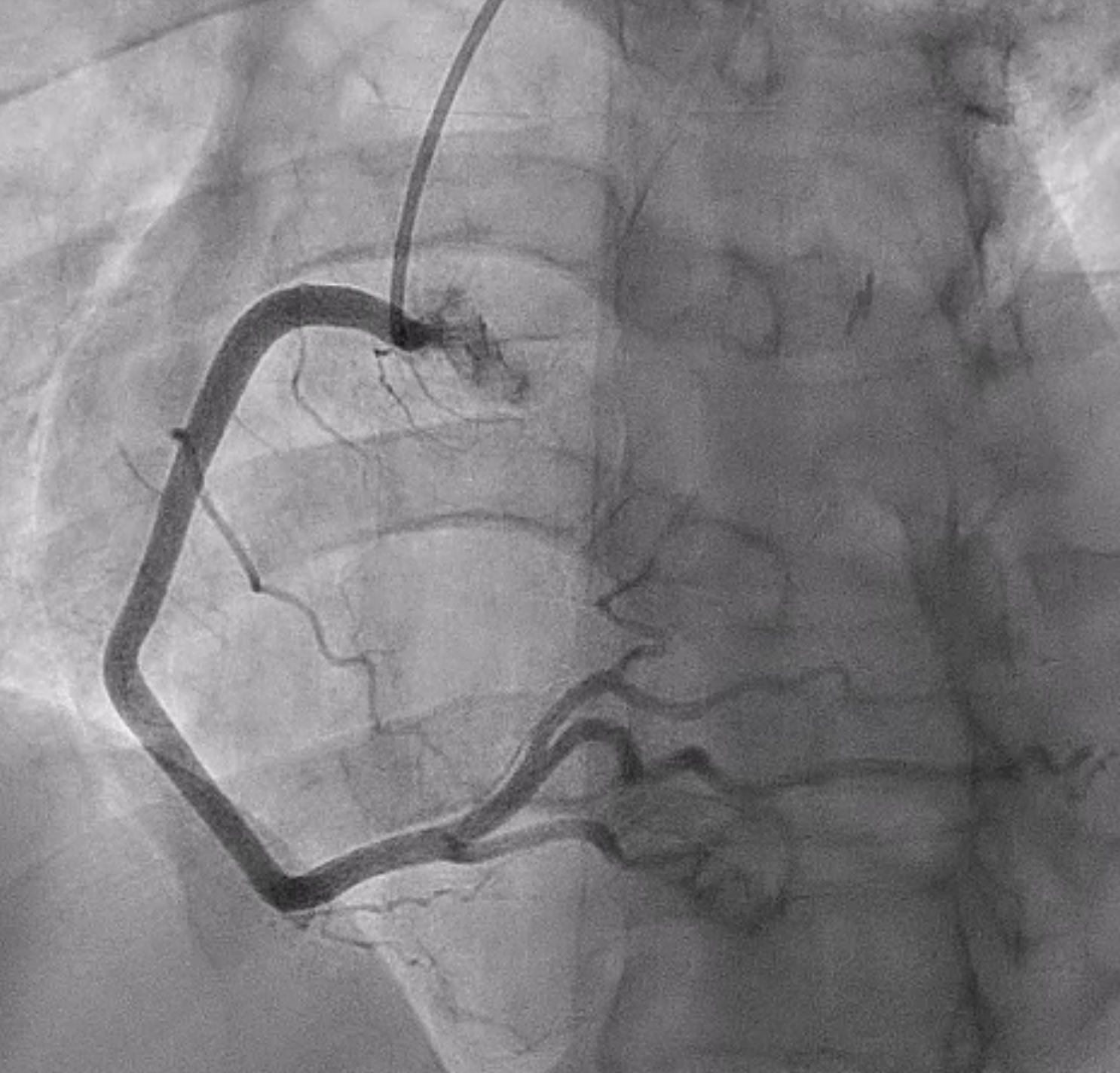
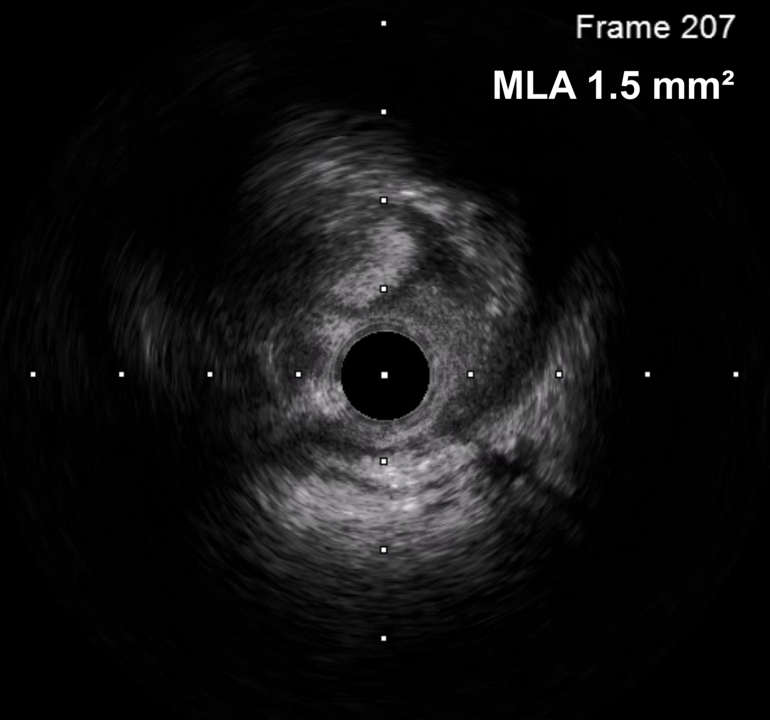
Patient had significant spasm of the bilateral radial arteries so right femoral artery access was obtained. A 6 Fr EBU 3.0 guide catheter was used to engage the left main coronary artery. A Sion wire was advanced into the LAD and a Sion Blue wire was advanced into the left circumflex. Pre-dilation was performed with a 2.5 x 12 mm semi-compliant balloon. IVUS showed MLA of 1.5 mm2 with intramural hematoma and evidence of plaque and thrombus. There was no evidence of SCAD on IVUS imaging. A 4 x 18 mm DES was placed in the left main and post-dilated with a 4.5 x 15 mm and 5 x 8 mm NC balloon. Final angiogram and IVUS confirmed excellent stent expansion/apposition with large MSA (11.8 mm2) and proximal stent edge in the ostium of the left main. Total air kerma was <500 mGy and and DAP was 50 Gy-cm2.
Pre-IVUS.mp4
Stent Position CRA25.mp4
Final Angiogram CAU27.mp4
Case Summary
Patient was started on dual antiplatelet therapy with aspirin and ticagrelor (non-responder to clopidogrel). She was also found to have 50-70% stenosis of the mid left internal carotid artery which is also likely attributed to her history of radiation therapy. Follow-up echo in 1 month showed normalization of her LVEF without wall motion abnormalities. The case highlights (1) pathophysiology of radiation induced coronary artery disease, and (2) minimizing radiation in pregnant patients needing complex coronary intervention.