
Lots of interesting abstracts and cases were submitted for TCTAP 2024. Below are the accepted ones after a thorough review by our official reviewers. Don’t miss the opportunity to expand your knowledge and interact with authors as well as virtual participants by sharing your opinion in the comment section!
TCTAP C-224
A Rare Giant Cardiac Squamous Cell Carcinoma in the Right Atrium : Timeline of Evidence
By Auliya Rezki Ananda, Az Hafid Nashar, Aussie Fitriani Ghaznawie, Jayarasti Kusumanegara, Richard Antonius
Presenter
Auliya Rezki Ananda
Authors
Auliya Rezki Ananda1, Az Hafid Nashar1, Aussie Fitriani Ghaznawie1, Jayarasti Kusumanegara1, Richard Antonius1
Affiliation
Hasanuddin University, Makassar, Indonesia1,
View Study Report
TCTAP C-224
Structural - Surgical Therapy (Structural)
A Rare Giant Cardiac Squamous Cell Carcinoma in the Right Atrium : Timeline of Evidence
Auliya Rezki Ananda1, Az Hafid Nashar1, Aussie Fitriani Ghaznawie1, Jayarasti Kusumanegara1, Richard Antonius1
Hasanuddin University, Makassar, Indonesia1,
Clinical Information
Patient initials or Identifier Number
Relevant Clinical History and Physical Exam
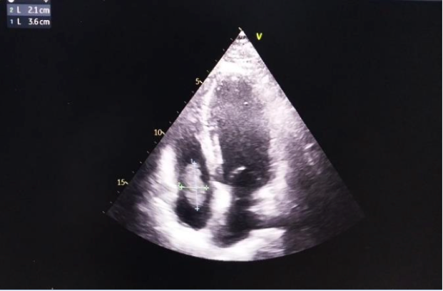
A 55-years-old male with shortness of breath over 2 months prior his admission to cardiac centre. He had no other cardiac manifestations before. Following his echocardiography, an osscilating mass sized 3.6 cm x 2.1 cm was stemming from his right atrium wall suggesting RA myxoma prolapsing toward his RV. He was then diagnosed with intracardiac mass suspected Cardiac Myxoma.
Relevant Test Results Prior to Catheterization
Relevant Catheterization Findings
Interventional Management
Procedural Step
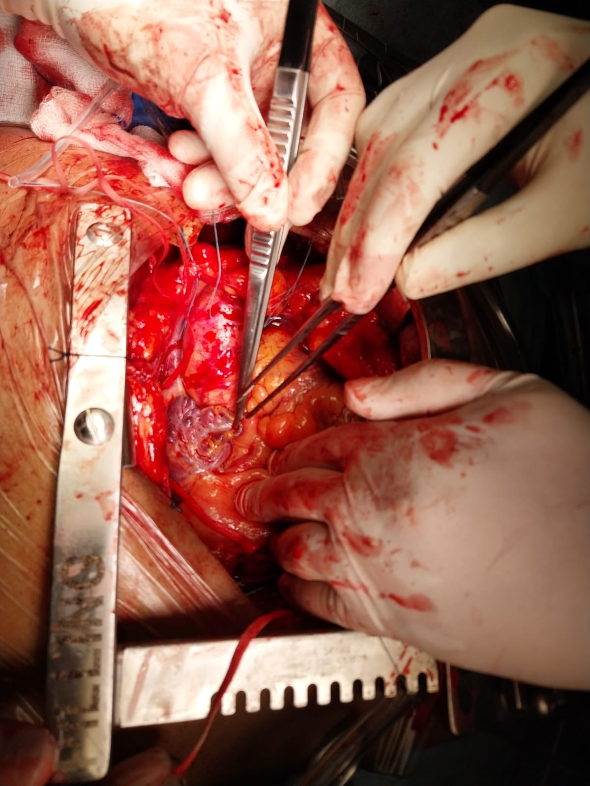
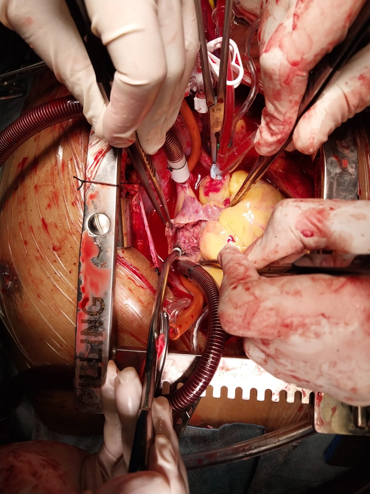
The patient was referred to Cardiac Surgeon to underwent open heart surgery for mass resection. An incision was made in the right atrium, a tumor mass size 4 x 2.8 x 0,8 cm was visible in the right atrial cavity, pink in color with a solid consistency and rough edges. The histopatology of the mass demonstrating a malignant tumor comes from squamous cell epithelium suggestive a metastatic squamous cell carcinoma. The post-operative course was uneventful.

The patient was suggested to undergo further investigations to assess the possible source of the primary tumor related to the cardiac mass but he refused to do another invasive examinations. Therefore tumor markers such CEA and Ca19-9 were checked. There is a significant increase in his CEA but his Ca 19-9 were in normal range.
Case Summary
The intracardiac mass in our patient was initially thought to be a cardiac myxoma, but the histopatology revealed a squamous cell carcinoma instead. The most common primary tumour found inside the heart cavity is cardiac myxoma. Though some complicated expressions have malignant characteristics, it is thought to be benign. The key components of a diagnostic workup include a high index of suspicion combined with a thorough and meticulous echocardiographic evaluation, followed by MDCT and/or CMR. To optimise the patient's benefit, the giant tumour was completely removed. Most importantly, echocardiography follow-up needs to be maintained to identify recurrence and long-term effects.