
Lots of interesting abstracts and cases were submitted for TCTAP 2024. Below are the accepted ones after a thorough review by our official reviewers. Don’t miss the opportunity to expand your knowledge and interact with authors as well as virtual participants by sharing your opinion in the comment section!
TCTAP C-040
A Case of In-Stent Intravascular Lithotripsy for Poor Stent Dilatation in a Severely Calcified Lesion
By Masahiro Shimoda, Hirohiko Ando, Akihiro Suzuki, Tetsuya Amano
Presenter
Masahiro Shimoda
Authors
Masahiro Shimoda1, Hirohiko Ando1, Akihiro Suzuki1, Tetsuya Amano1
Affiliation
Aichi Medical University, Japan1,
View Study Report
TCTAP C-040
Coronary - Complex PCI - Calcified Lesion
A Case of In-Stent Intravascular Lithotripsy for Poor Stent Dilatation in a Severely Calcified Lesion
Masahiro Shimoda1, Hirohiko Ando1, Akihiro Suzuki1, Tetsuya Amano1
Aichi Medical University, Japan1,
Clinical Information
Patient initials or Identifier Number
Relevant Clinical History and Physical Exam
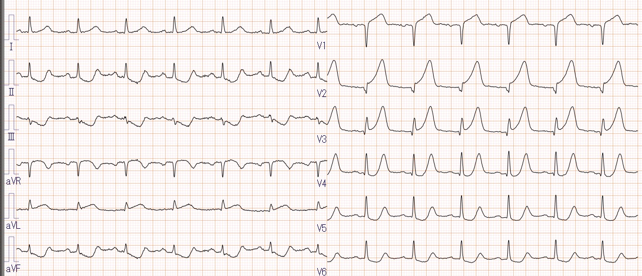
A 74-year-old female presented with chest pain lasting 2 hours prior to hospital admission. Upon her arrival at the hospital, the patient still reported chest symptoms. An electrocardiogram revealed ST-segment elevation in V2-5, I, and aVL, and transthoracic echocardiography showed a decrease in anteroseptal wall motion.
Relevant Test Results Prior to Catheterization
Relevant Catheterization Findings
Emergency coronary angiography demonstrated a total occluded lesion with severe calcification and the presence of a thrombus in the proximal anterior descending artery. The lesion was pretreated with a 2.5 x 15 mm non-compliant balloon and a 2.75 x 15 mm cutting balloon, followed by placement of a 2.75 x 28 mm Synergy stent. After stent placement, post-dilatation was performed with a 3.0 x 12 mm non-compliant balloon, but intravascular ultrasound (IVUS) imaging showed insufficient stent dilat
Interventional Management
Procedural Step
Emergency coronary angiography demonstrated a total occluded lesion with severe calcification and the presence of a thrombus in the proximal anterior descending artery. The lesion was pretreated with a 2.5 x 15 mm non-compliant balloon and a 2.75 x 15 mm cutting balloon, followed by placement of a 2.75 x 28 mm Synergy stent. After stent placement, post-dilatation was performed with a 3.0 x 12 mm non-compliant balloon, but intravascular ultrasound (IVUS) imaging showed insufficient stent dilatation due to calcification (minimal stent area; 2.8mm2). Consequently, a 3.0 x 12 mm IVL was performed within the stented segment, and additional dilation was performed with a 3.5 x 12 mm non-compliant balloon. The final IVUS image showed effective stent dilatation with a minimal stent area of 5.5 mm2.

Case Summary
Intravascular lithotripsy (IVL) is a novel approach for treating de novo calcified coronary lesions, which improves procedural outcomes. However, there is limited data on the use of IVL in stent-implanted lesions.The present case highlights the effectiveness of IVL for lesions with stent under expansion just after stenting. There have been anecdotal reports of stent polymer damage when IVL is performed inside the stent, however such concerns were unnecessary in this case because the stent used was a drug-eluting stent with an abluminal coating. We believe this case provides valuable insights into this area of inquiry.