
Lots of interesting abstracts and cases were submitted for TCTAP 2024. Below are the accepted ones after a thorough review by our official reviewers. Don’t miss the opportunity to expand your knowledge and interact with authors as well as virtual participants by sharing your opinion in the comment section!
A Challenging Case: What Is the Cause of Shock?
By Ricky Wang Hei Leung
Presenter
Ricky Wang Hei Leung
Authors
Ricky Wang Hei Leung1
Affiliation
Queen Mary Hospital, Hong Kong, China1,
View Study Report
Coronary - Complication Management
A Challenging Case: What Is the Cause of Shock?
Ricky Wang Hei Leung1
Queen Mary Hospital, Hong Kong, China1,
Clinical Information
Patient initials or Identifier Number
Relevant Clinical History and Physical Exam
Our case was an 82-year-old lady who had a background of hypertension and diabetes mellitus on insulin. She was initially admitted to the hospital for class III anginal symptoms. A CT coronary angiogram was performed showing triple vessel disease with extensive atherosclerotic changes. Thus an early elective PCI was arranged.
Physical examination was otherwise unremarkable.
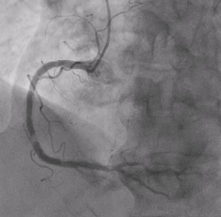
On coronary angiogram, there were multiple critical calcified stenoses over the RCA.
Physical examination was otherwise unremarkable.
On coronary angiogram, there were multiple critical calcified stenoses over the RCA.
Relevant Test Results Prior to Catheterization
A CT coronary angiogram was performed showing triple vessel disease with extensive atherosclerotic changes.
Relevant Catheterization Findings
Coronary angiogram showed calcified vessels. Left sided disease was mild to moderate in severity. However, the dominant RCA showed multiple critical calcified stenosis.
IVUS could not cross the calcified lesion. Rotablation followed by IVL (Shockwave) were performed. Temporary pacing lead was also inserted before rotablation. RCA was later treated with 4 stents.
However, the patient suffered from shock after stent implant.
IVUS could not cross the calcified lesion. Rotablation followed by IVL (Shockwave) were performed. Temporary pacing lead was also inserted before rotablation. RCA was later treated with 4 stents.
However, the patient suffered from shock after stent implant.
Interventional Management
Procedural Step
On assessment of shock, patient was volume depleted.
An abdominal aortogram showed contrast extravasation at Rt inferior epigastric artery and branches of internal iliac artery likely due to vascular injury during RFV puncture for temporary pacing lead insertion.
We immediately established contralateral (i.e., left) access and did selective injection to identify bleeding sites and balloon tamponade to support the hemodynamics of the patient.
Finally, hemostasis was achieved by coil embolisation and covered stent implantation.
Case Summary
There are several causes of shock in complex PCI. Identifying the cause of shock is of paramount importance so that effective treatment, not only treatment of stabilizing the patient's hemodynamics but also treatment of complications if they occur, can be established. In this case, vessel injury was caused by multiple attempts of blind puncture of RFV, leading to bleeding complications. Two ways of treatment of vessel perforation i.e., coil embolization and cover stent were demonstrated.