
Lots of interesting abstracts and cases were submitted for TCTAP 2024. Below are the accepted ones after a thorough review by our official reviewers. Don’t miss the opportunity to expand your knowledge and interact with authors as well as virtual participants by sharing your opinion in the comment section!
TCTAP C-181
Unexpected Intramural Hematoma in Optical Coherence Tomography Guided Percutaneous Coronary Intervention
By Masahiro Katamine, Yosuke Fujiki, Tsuyoshi Nozue, Ichiro Michishita
Presenter
Masahiro Katamine
Authors
Masahiro Katamine1, Yosuke Fujiki1, Tsuyoshi Nozue2, Ichiro Michishita1
Affiliation
Yokohama Sakae Kyosai Hospital, Japan1, , Japan2,
View Study Report
TCTAP C-181
Coronary - Imaging & Physiology - Invasive Imaging (IVUS, OCT, NIRS, VH, etc)
Unexpected Intramural Hematoma in Optical Coherence Tomography Guided Percutaneous Coronary Intervention
Masahiro Katamine1, Yosuke Fujiki1, Tsuyoshi Nozue2, Ichiro Michishita1
Yokohama Sakae Kyosai Hospital, Japan1, , Japan2,
Clinical Information
Patient initials or Identifier Number
Relevant Clinical History and Physical Exam
A81-year-old woman was referred to our hospital suffering from chest pain and impaired exercise tolerance for 1 month. She had cardiovascular risk factors such as dyslipidemia and hypertension. Her physical examination was normal.
Relevant Test Results Prior to Catheterization
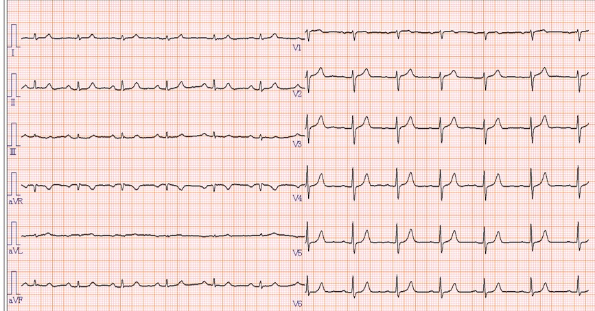
There were no remarkable findings in laboratory tests, electrocardiography and echocardiography. Coronary computed tomography showed a moderate stenosis in both right coronary artery (RCA) and left anterior descending artery (LAD).
Relevant Catheterization Findings
A coronary angiogram showed a severe stenosis at mid RCA (Panel A) and a moderate stenosis at proximal LAD.

Interventional Management
Procedural Step
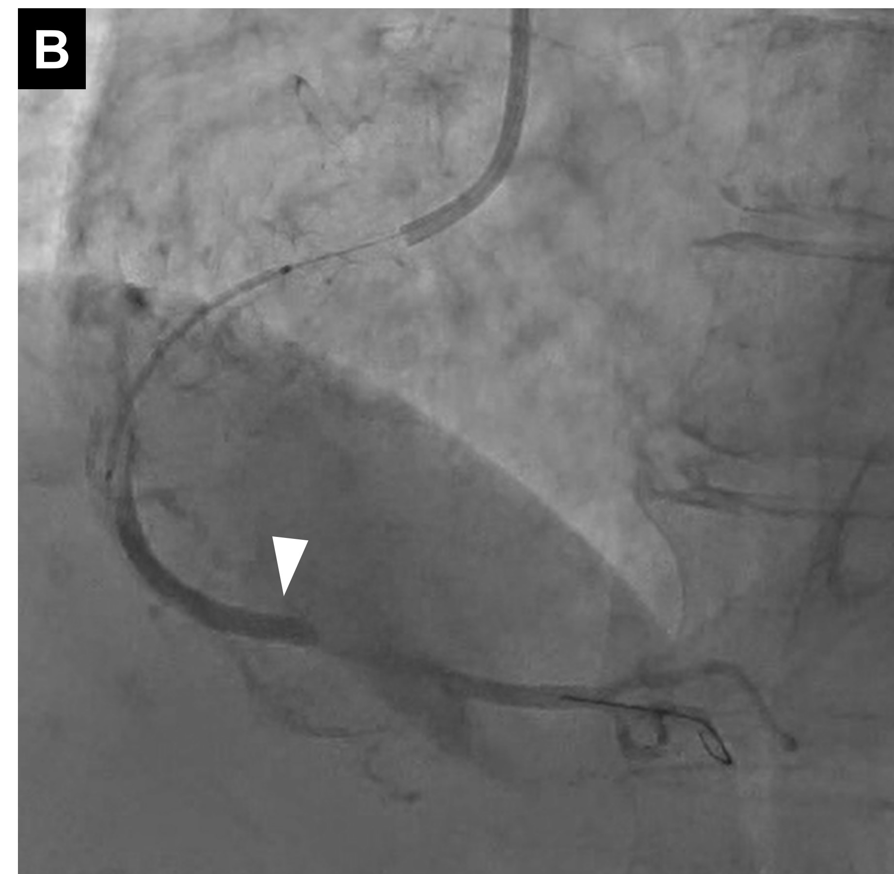
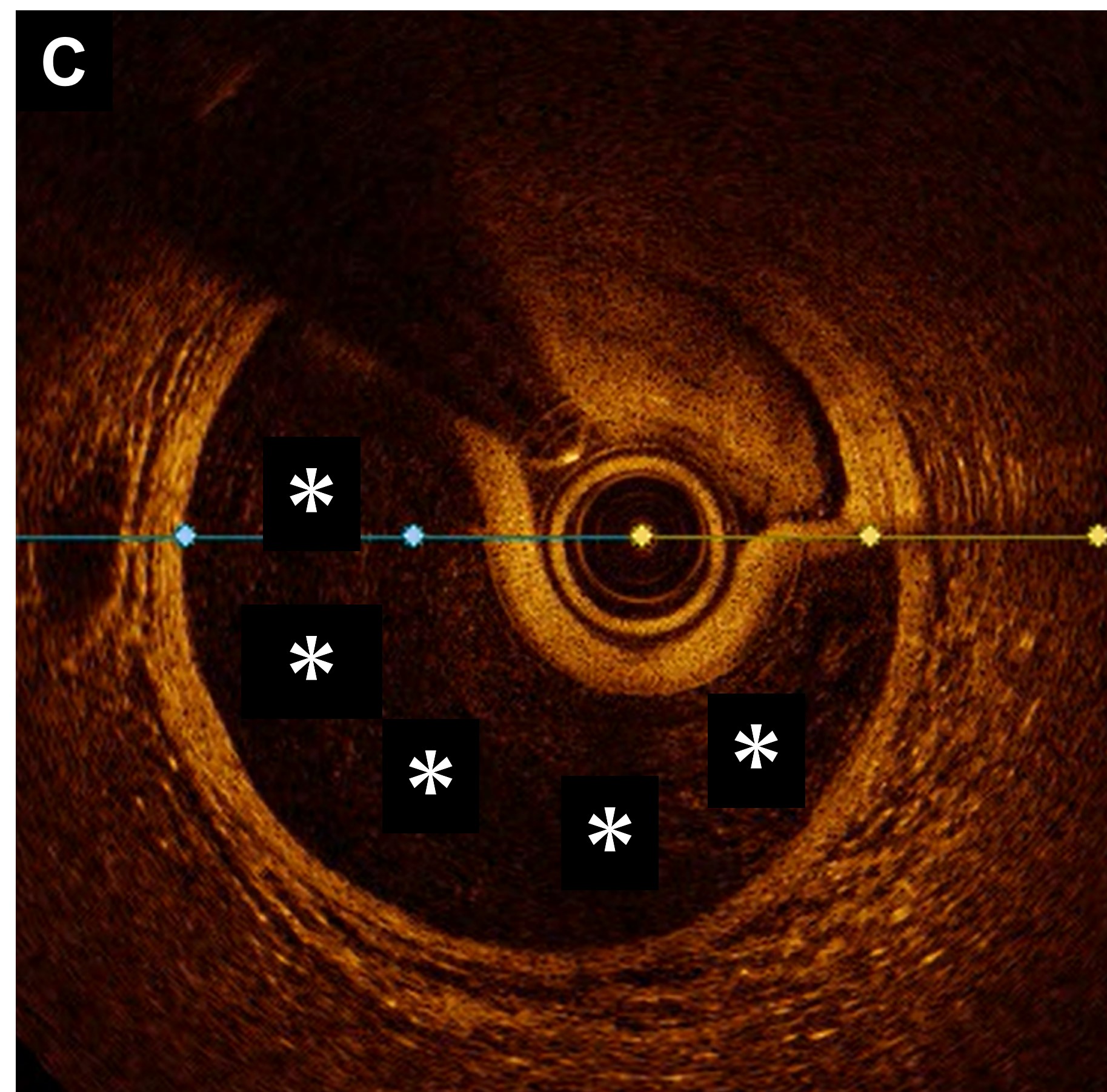
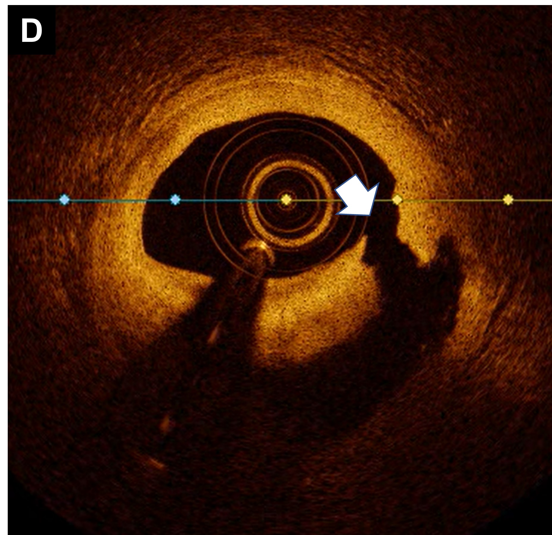
She underwent percutaneous coronary intervention (PCI) for a focal lesion at mid RCA. Pre-dilatation was performed using a small balloon with 2.25mm in diameter, because antegrade coronary flow was insufficient to enable optical coherence tomography (OCT). There appeared to be a propagating the coronary dissection on angiogram by the contrast injection just before stent implantation. Angiography demonstrated the contrast pooled in distal RCA (Panel B). OCT showed circumferential coronary hematoma at the distal of RCA (Panel C). Looking back at the first OCT, a significant medial dissection of the location performed pre-dilatation was observed (Panel D). Two zotarolimus-eluting stents implantation successfully compressed the hematoma.
Case Summary
We report an unexpected intramural hematoma in OCT-guided PCI due to pre-dilatation with a small balloon. The contrast flush might have generated a propagating the dissection and expanding an intramural hematoma, because a medial dissection due to pre-dilatation was observed by first OCT. Although OCT has an excellent imaging modality, physicians should remember that pre-dilatation to enable OCT imaging can cause intramural hematoma by contrast flush during PCI.