
Lots of interesting abstracts and cases were submitted for TCTAP 2024. Below are the accepted ones after a thorough review by our official reviewers. Don’t miss the opportunity to expand your knowledge and interact with authors as well as virtual participants by sharing your opinion in the comment section!
TCTAP C-047
A Calcified Left Main Intervention With Lithotripsy and Intravascular Imaging
By Ranjan Modi
Presenter
Ranjan Modi
Authors
Ranjan Modi1
Affiliation
Sarvodaya Healthcare, India1,
View Study Report
TCTAP C-047
Coronary - Complex PCI - Calcified Lesion
A Calcified Left Main Intervention With Lithotripsy and Intravascular Imaging
Ranjan Modi1
Sarvodaya Healthcare, India1,
Clinical Information
Patient initials or Identifier Number
Relevant Clinical History and Physical Exam
A 57 year old male , HbsAg positive with history of hypertension presented with complaints of chest pain NYHA II- III associated with dyspnoea. on Examination: BP: 148/86mmHg , HR: 82/ min , regular , CVS and R/S : Nothing significant.
Relevant Test Results Prior to Catheterization
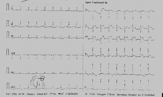
ECG : ST- T changes in anterior leads with T wave inversionsEcho : Hypokinesia of LAD territory with LVEF 40%

Relevant Catheterization Findings
CAG : Left dominant sysytem Left Main Distal 80-90% , severely calcified LAD ostial 80-90% severely calcified proximal long segment 80% with mid LAD 70% lCX : Dominant . ostial : mild plaque , proximal and mid : mild plaque
Interventional Management
Procedural Step
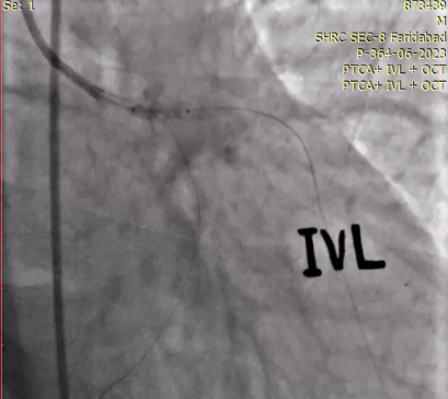
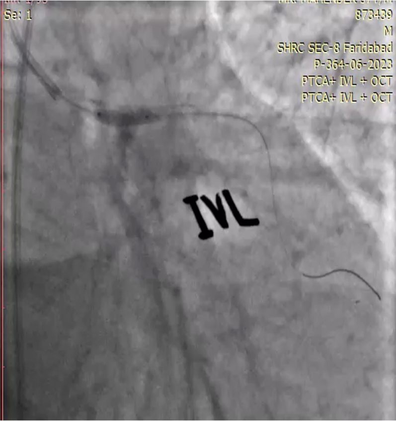
LEFT CORONARY WAS CATHETERISED USING XB 3.5 7 F GUIDING . FLOPPY WIRES CROSSED INTO LAD AND LCX. PRE DILATATION DONE USING 2X 12 AND 3X12 NC BALLON IN MID, OSTIAL LAD AND DISTAL LEFT MAIN. OCT RUN TAKEN FROM LEFT MAIN TO LAD SHOWED SEVERE CALCIFIED DISTAL LEFT MAIN AND OSTIAL LAD WITH A LONG SEGMENT DISEASE IN LAD . IVL DONE USING 3X 12 BALLOON WITH 5 CYCLES OF IVL. DES IMPLANTED FROM LEFT MAIN TO LAD 3X32 . POST DILATATION DONE USING 3X10 NC BALLON IN LAD AND 3.5 X 10 NC BALLON IN LM AND OSTIAL LAD. CHECK ANGIO SHOWED RESIDUAL CALCIUM IN DISTAL LEFT MAIN WITH UNDEREXPANDED STENT IN DISTA LEFT MAIN AND OSTIAL LAD. REPEAT IVL DONE USING 3X12 BALLON AND REMAINING 3 CYCLES . OCT RUN SHOWED UNDEREXPANDED STENT . POST DILATTION DONE USING 4X10 NC BALLOON IN LEFT MAIN . FINAL RESULT IS SATISFACTORY WITH TIMI III FLOW , NO DISSECTION , NO THROMBUS AND WELL EXPANDED STENT .

Case Summary
Calcified left main intervention is an arduous and challenging procedure.
Tackling coronary calcium is important to optimize the stent deployment and improve the long term outcomes.
Intracoronary imaging is a Class I indication for left main interventions.
Pre PTCA OCT run helps to identify the lesion characteristics , calcium burden and procedural planning.
Post PTCA OCT evaluates the adequate stent deployment and help stent apposition.
All calcified coronary interventions should be attempted using debulking devices and imaging to improve cardiovascular mortality and mortality.
IVL has significant advantages over other calcium debulking devices like
Maintaining a wire in the side branch.
Repetitive use of the device even after stent deployment.
Ease of use of IVL with practically minimum learning curve.