
Lots of interesting abstracts and cases were submitted for TCTAP 2023. Below are the accepted ones after a thorough review by our official reviewers. Don’t miss the opportunity to expand your knowledge and interact with authors as well as virtual participants by sharing your opinion in the comment section!
TCTAP C-155
A Case Report: Successful Removal of an Entrapped Stent Delivery System With an Additional Access
By Eiji Miyauchi
Presenter
Eiji Miyauchi
Authors
Eiji Miyauchi1
Affiliation
Kagoshima City Hospital, Japan1,
View Study Report
TCTAP C-155
ENDOVASCULAR - Peripheral Vascular Disease and Intervention
A Case Report: Successful Removal of an Entrapped Stent Delivery System With an Additional Access
Eiji Miyauchi1
Kagoshima City Hospital, Japan1,
Clinical Information
Patient initials or Identifier Number
K.T.
Relevant Clinical History and Physical Exam
A 70-year-old man with a history of hypertension and cerebral infarction presented to our hospital with a chief complaint of rest pain in the left lower limb and a right foot ulcer. Both feet were cold and cyanotic. The popliteal, dorsalis pedis, or posterior tibial artery in his right lower limb was not palpable. The common femoral, popliteal, dorsalis pedis, or posterior tibial artery in his left lower limb was also not palpable.
Relevant Test Results Prior to Catheterization
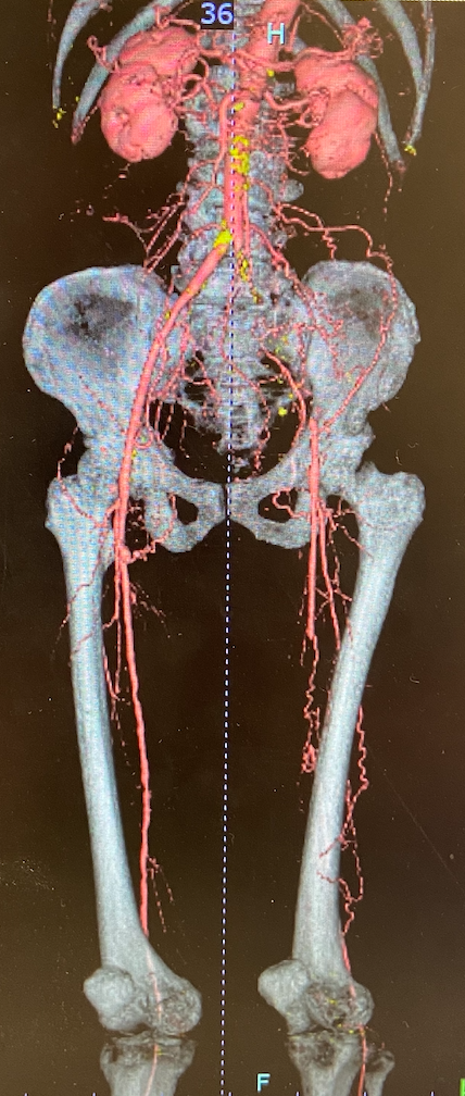
His ankle-brachial index was 0.61/error. Computed tomography angiography showed complete occlusion of the right superficial femoral artery (SFA), complete occlusion of the left common iliac artery (CIA) to the external iliac artery (EIA), and complete occlusion of the left superficial femoral artery. First, endovascular treatment (EVT) to the iliac artery region was performed without complication.
Relevant Catheterization Findings
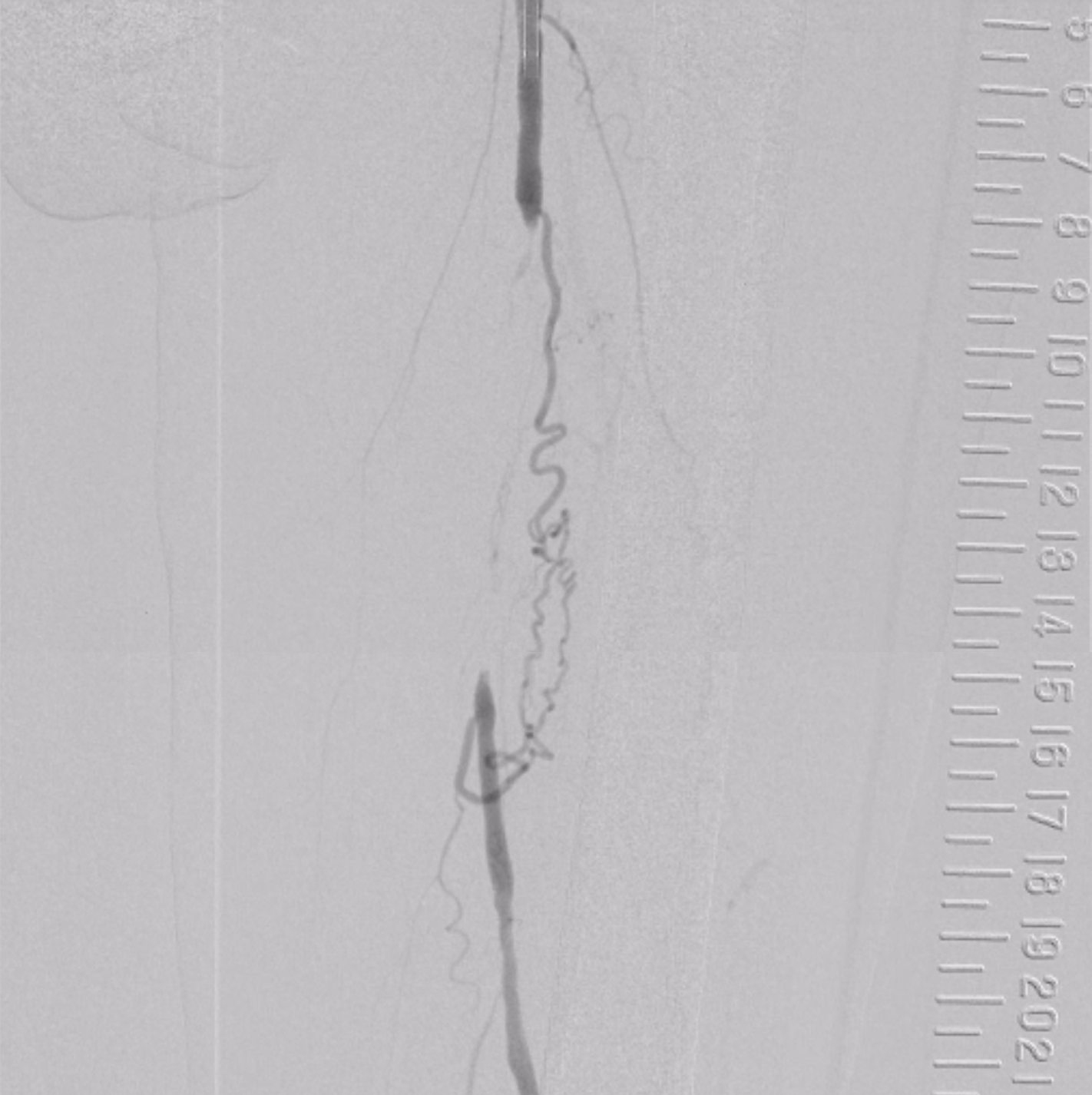
First, endovascular treatment (EVT) to the iliac artery region was performed without complication. At a later date, EVT to the left SFA was performed. Initial angiography showed a completely occluded lesion with an occlusion length of approximately 12 cm in the middle portion and localized severe stenosis distal to the SFA. The overall lesion length was 20 cm. There was no significant calcification throughout the lesion. There was no significant stenosis peripherally from the popliteal artery.
Interventional Management
Procedural Step
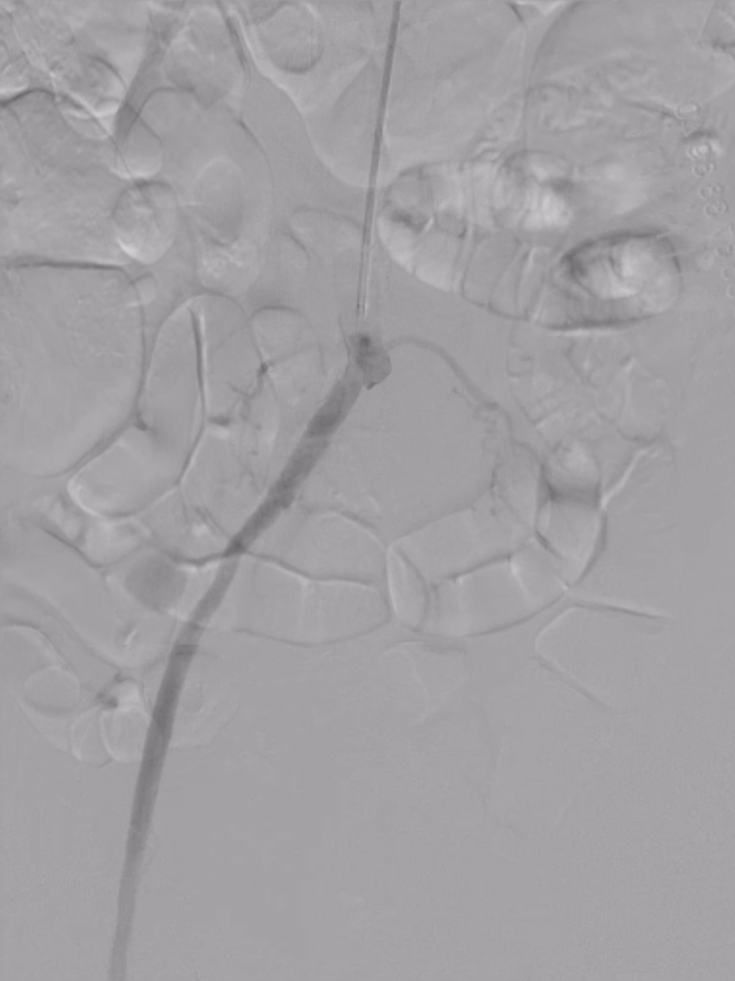
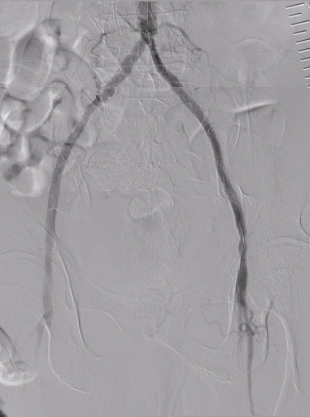
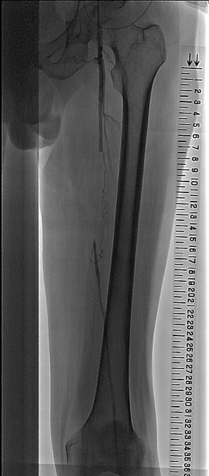
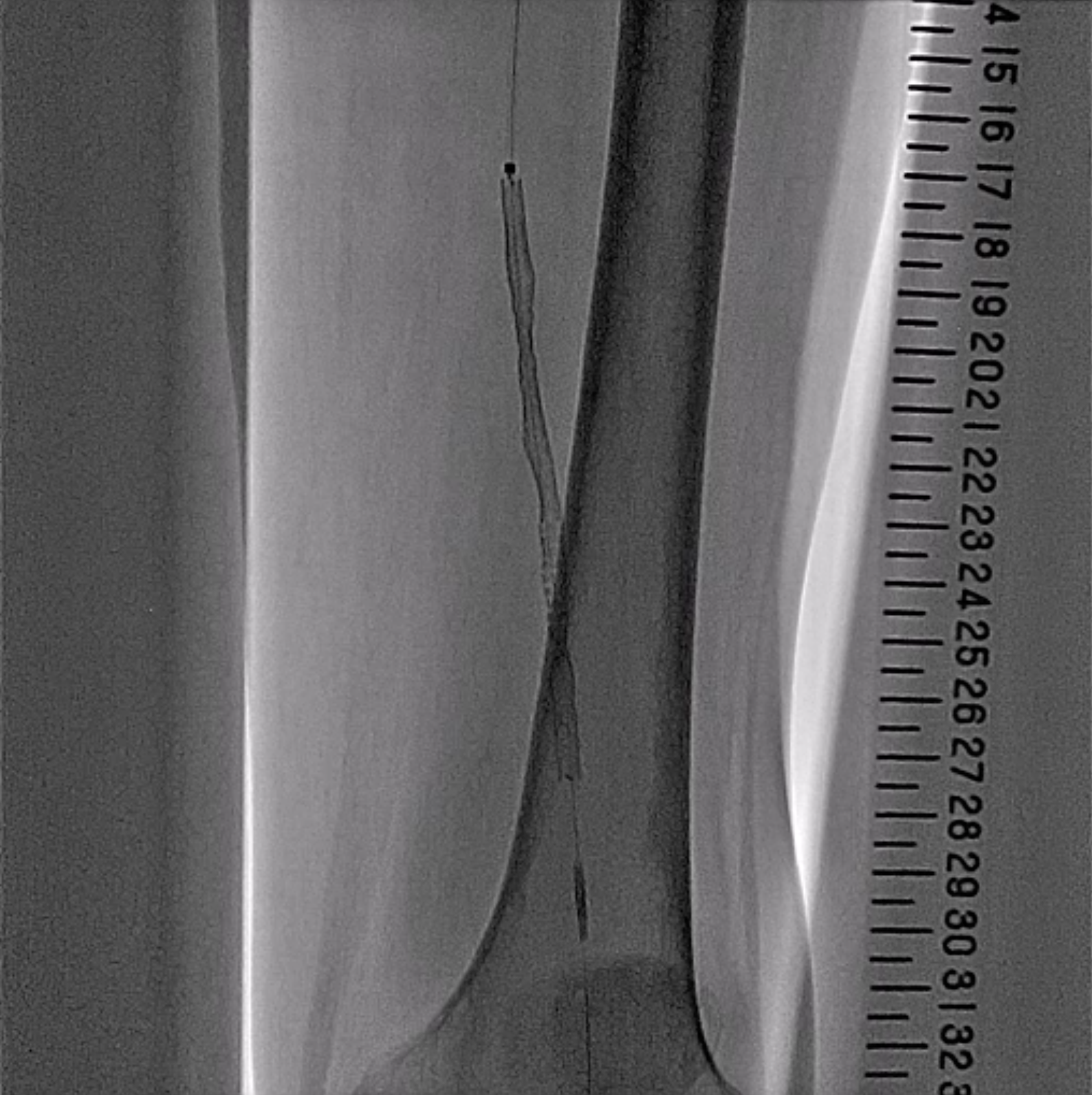
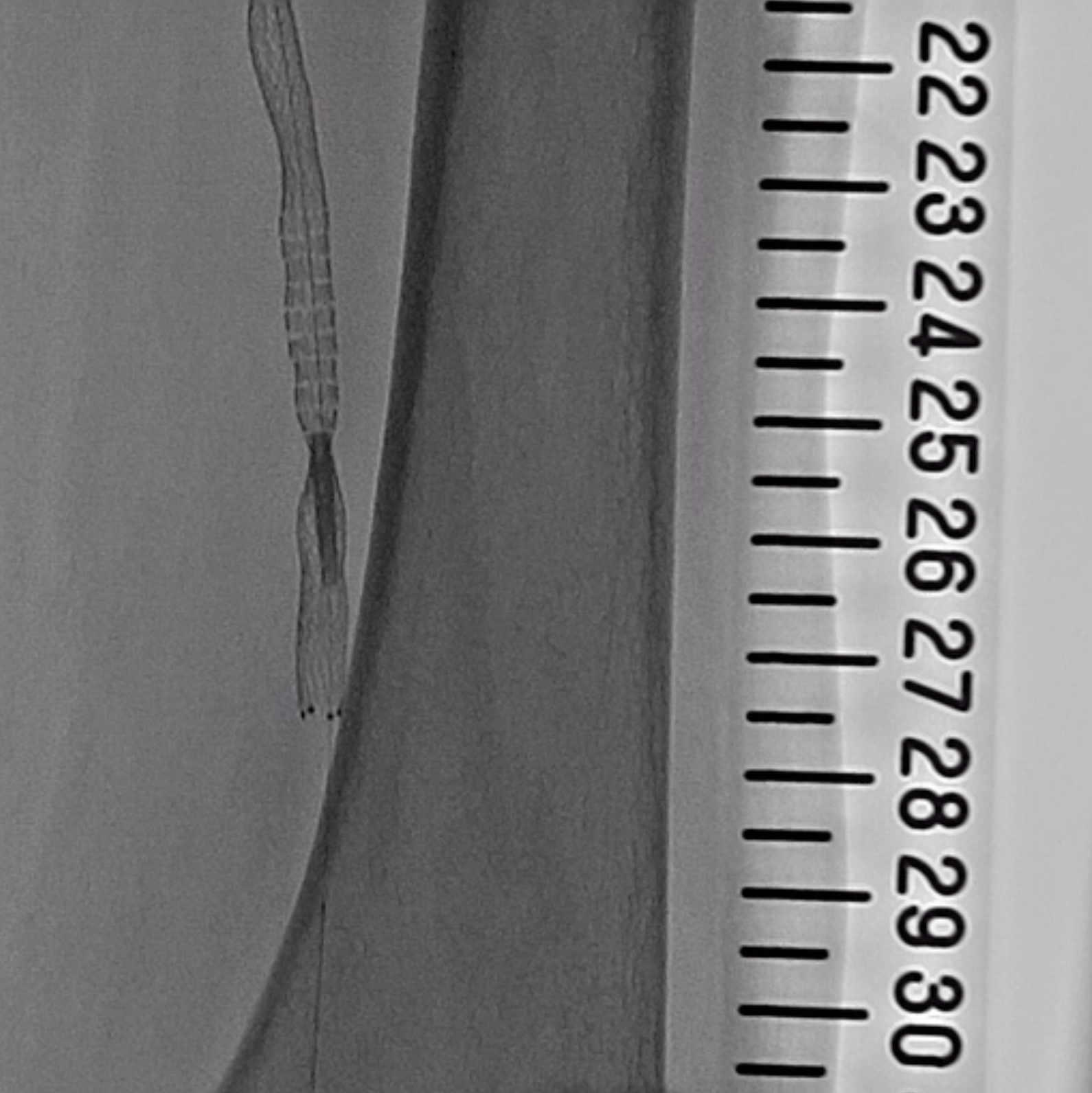
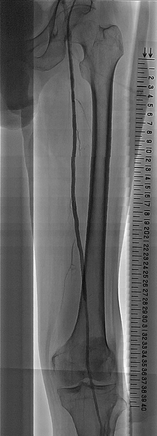
A 6 Fr Parent Plus 23 cm (Medikit, Japan) was inserted antegrade into the left common femoral artery. 0.014 Jupitar FC 235 cm (Boston, USA) was used for wiring with the support of Corsair PV 135 cm (ASAHI intec, Japan). The guidewire stopped advancing in the middle of the lesion and was changed to 0.014 Astato XS 9-12 180 cm (ASAHI intec, Japan) and successfully passed through the lesion. The occluded lesion was dilated with a Coyote 4.0 / 150 mm (Boston, USA). IVUS revealed severe localized stenosis distal to the SFA (25 cm on the scale), followed by moderate stenosis. An ELUVIA 6.0/120 mm (Boston, USA) was implanted to cover all stenotic lesions. The stent was inadequately dilated at the 25 cm site on the scale. The tip of the stent delivery system was caught in the poorly dilated site and could not be removed. 4 Fr sheath was inserted antegrade into the left common femoral artery, and the poorly dilated site was passed through the stent with 0.014 Command 250 cm (Abbot, USA). The poorly dilated site was dilated with Coyote 3.0 /20 mm (Boston, USA). The indentation of the site was resolved. As a result, the stent delivery system was successfully removed. After that, ELUVIA 6.0 / 120 mm (Boston, USA) was implanted to cover the occluded lesion. Additional in-stent dilation with Coyote 4.0 / 150 mm was added and stenosis improved to 0 %. Two puncture sites were successfully stopped by manual compression.
Case Summary
Failure to perform adequate pre-dilation led to poor stent dilation, which resulted in difficulty in removing the stent system. Forced removal was considered to cause stent fracture. Although it increased the number of puncture sites, we selected the minimum sheath size, dilated the poorly dilated site sufficiently, and safely removed the stent delivery system. We consider this case to be a lesson to be learned and report it here.