
Lots of interesting abstracts and cases were submitted for TCTAP 2023. Below are the accepted ones after a thorough review by our official reviewers. Don’t miss the opportunity to expand your knowledge and interact with authors as well as virtual participants by sharing your opinion in the comment section!
TCTAP A-059
Comparison of Contrast Media Versus Hydroxyethyl Starch for Frequency-Domain Optical Coherence Tomography Imaging in Coronary Artery Disease
By Dong Oh Kang, Hyeong Soo Nam, Sunwon Kim, Hongki Yoo, Jin Won Kim
Presenter
Dong Oh Kang
Authors
Dong Oh Kang1, Hyeong Soo Nam2, Sunwon Kim3, Hongki Yoo2, Jin Won Kim1
Affiliation
Korea University Guro Hospital, Korea (Republic of)1, KAIST, Korea (Republic of)2, Korea University Ansan Hospital, Korea (Republic of)3
View Study Report
TCTAP A-059
Imaging: Intravascular
Comparison of Contrast Media Versus Hydroxyethyl Starch for Frequency-Domain Optical Coherence Tomography Imaging in Coronary Artery Disease
Dong Oh Kang1, Hyeong Soo Nam2, Sunwon Kim3, Hongki Yoo2, Jin Won Kim1
Korea University Guro Hospital, Korea (Republic of)1, KAIST, Korea (Republic of)2, Korea University Ansan Hospital, Korea (Republic of)3
Background
Contemporary high-resolution intracoronary optical coherence tomography (OCT) requires injection of flushing media for image acquisition to avoid signal attenuation by red blood cells. There is a growing need for an alternative flushing media other than iodine contrast to reduce the risk of renal dysfunction. To address this issue, we investigated the safety and feasibility of hydroxyethyl starch (HES) as a flushing media for OCT image acquisition and compared with the contrast media flushed images.
Methods
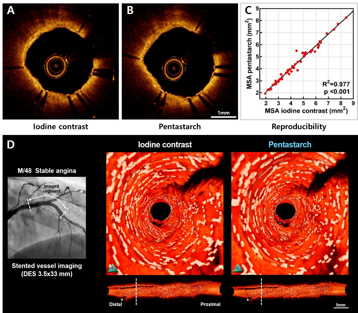
We prospectively enrolled 43 patients with 70 coronary lesions (46 stented and 24 pre-stented or non-culprit lesions). Paired OCT images were obtained by manual injection of iodine contrast (average 12 mL),followed by pentastarch (10% HES 260 kDa/0.45, average 15 mL). A total of 162 runs(81 pairs) were analyzed and each OCT pullback was assessed on a frame-by-frame basis by automated customized lumen contour and stent strut segmentation algorithm. The number of clear image segments (CIS) and pre-specified quantitative 2D- and 3D-morphometric measurements were compared between the paired OCT images.
Results
Image quality as assessed by proportion of CIS were comparable between the contrast media and pentastarch flushed images (93.3%vs. 93.7%; p=0.501). Pixel-based blood flushing capability showed no significant difference between the paired image segments (median (IQR), 0.951 (0.947-0.953)vs. 0.951 (0.949-0.952), p=0.264; Figure A-B). Quantitative 2D- and 3D-morphometric measurements of minimal stent area, minimal lumen area, segment stent volume and segment lumen volume correlated well between the paired OCT runs (all p<0.001; Figure C). Customized 3D-rendered reconstruction of paired OCT images enabled clear visualization of stent strut geometry within the stented vessels (Figure D). There were no clinically relevant complications associated with OCT imaging with pentastarch administration such as anaphylactic reaction, life-threatening arrhythmia, heart failure, or acute renal deterioration.
Conclusion
Intracoronary OCT using pentastarch is safe and technically feasible without affecting the optical signal intensity and overall image quality. Non-contrast imaging with pentastarch significantly reduces the obligatory iodine contrast load, therefore, could be considered as a promising strategy in high-risk patients for renal impairment.