Lots of interesting abstracts and cases were submitted for TCTAP & AP VALVES 2020 Virtual. Below are accepted ones after thoroughly reviewed by our official reviewers. Don’t miss the opportunity to explore your knowledge and interact with authors as well as virtual participants by sharing your opinion!
* The E-Science Station is well-optimized for PC.
We highly recommend you use a desktop computer or laptop to browse E-posters.
CASE20191026_003
| CORONARY - Acute Coronary Syndromes (STEMI, NSTE-ACS) | |
| Paradoxical Coronary Artery Embolism with Decreased Protein C Activity | |
| Emiko Oishi1 | |
| Osaka Saiseikai Izuo Hospital, Japan1, | |
|
[Clinical Information]
- Patient initials or identifier number:
K.K
-Relevant clinical history and physical exam:
This is 30’s old male without any past history of illness. He had no past history of Kawasaki disease. He had no family history of ischemic heart disease. His coronary risk factor was smoking. He came to our hospital on foot for sudden onset of chest pain.
-Relevant test results prior to catheterization:
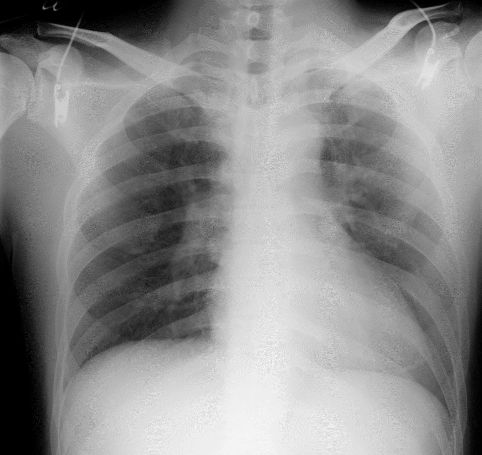
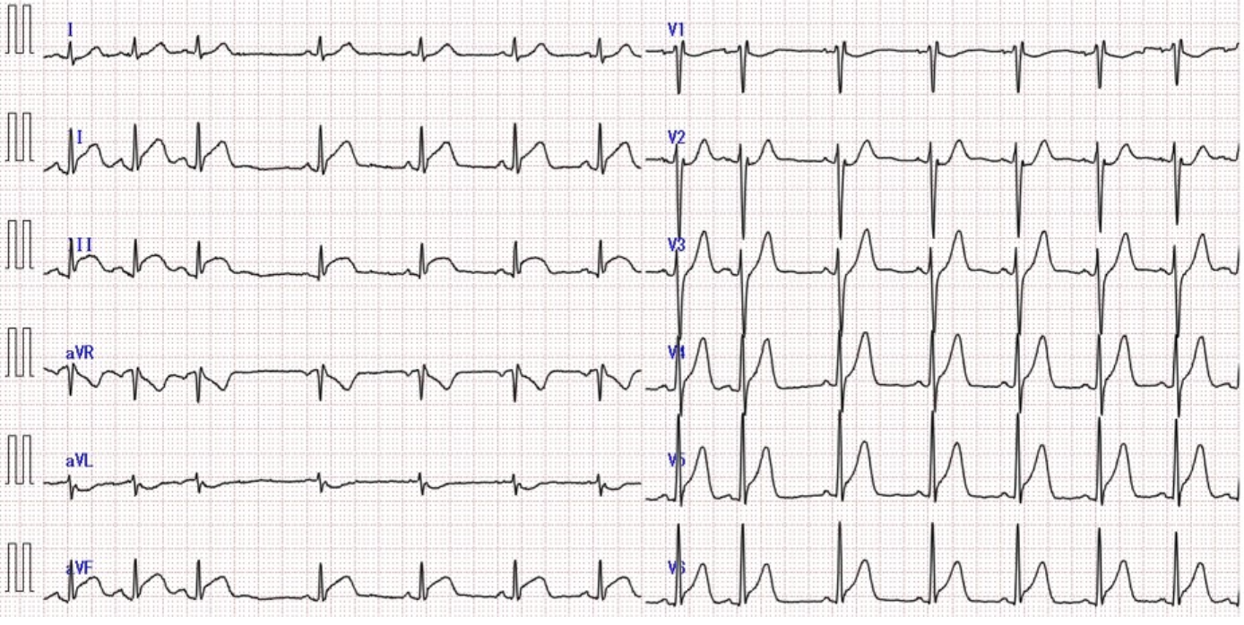
His ECG showed normal sinus rhythm , ST elevationin in Ⅱ, Ⅲ, aVF, V4-6. His chest X-ray showed cardiomegaly and lung congestion. His blood test resulted WBC was increased but cardiac enzyme was not elevated at that time. We diagnosed him STEMI. We performed emergency coronary angiogram (CAG).
  - Relevant catheterization findings:
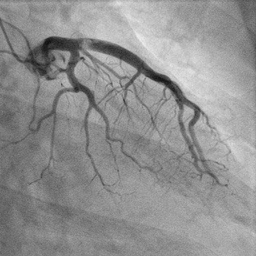
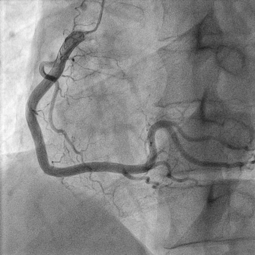
It showed there is a big thrombus at proximal LAD, total occlusion with thrombus in distal LAD without any atherosclerotic changes. He underwent primary PCI to LAD.
  |
|
|
[Interventional Management]
- Procedural step:
We aspirated thrombus by the Ribirth aspiration catheter. We could get TIMI-3 flow after thrombus aspiration, but the thrombus was still existed at the proximal LAD. We finished this procedure because of got TIMI-3 flow.We performed CAG and OCT 3days after primary PCI to check thrombus. His LAD had TIMI-3 flow and the existed thrombus was disappeared from angiogram. The OCT image showed a mural thrombus at the proximal LAD without ruptured plaque. His coronary CT also showed mural thrombus at the proximal LAD. We suspected the thrombus came from the extra heart. His protein C activity was decreased by his blood test. His bubble test by the trans esophageal echocardiography was grade3 positive. We diagnosed him a paradoxical coronary artery embolism with decreased protein C activity. His OCT findings and coronary CT findings showed the thrombus was organized.
- Case Summary:
We experienced STEMI in young adult. The cause of this ACS suspected extra heart thrombus. We diagnosed him a paradoxical coronary artery embolism with decreased protein C activity and existed patent foramen ovale. It is important to investigate the cause of ACS especially for a young adult to determination of treatment policy.
|
|