E-Case

JACC

Lots of interesting abstracts and cases were submitted for TCTAP 2025. Below are the accepted ones after a thorough review by our official reviewers. Don¡¯t miss the opportunity to expand your knowledge and interact with authors as well as virtual participants by sharing your opinion in the comment section!

TCTAP C-120

Endless Loops: The Infinite Challenge of Complex RCA CTO Intervention

By Kai Soon Liew, Huan Yean Kang, Chai Yih Tan, Virginia Nancy, Kay Leong Kenneth Khoo, Subramaniam Prabahkar, Mohd Khairi Othman, Narasamuloo Kantha Rao, Krishinan Saravanan, Chee Tat Liew, Karthikesan Dharmaraj

Presenter

Chai Yih Tan

Authors

Kai Soon Liew1, Huan Yean Kang1, Chai Yih Tan1, Virginia Nancy1, Kay Leong Kenneth Khoo1, Subramaniam Prabahkar1, Mohd Khairi Othman1, Narasamuloo Kantha Rao1, Krishinan Saravanan1, Chee Tat Liew2, Karthikesan Dharmaraj1

Affiliation

Hospital Sultanah Bahiyah, Malaysia1, Penang Pantai Hospital, Malaysia2,
View Study Report
TCTAP C-120
Coronary - Complex PCI - Long Lesion

Endless Loops: The Infinite Challenge of Complex RCA CTO Intervention

Kai Soon Liew1, Huan Yean Kang1, Chai Yih Tan1, Virginia Nancy1, Kay Leong Kenneth Khoo1, Subramaniam Prabahkar1, Mohd Khairi Othman1, Narasamuloo Kantha Rao1, Krishinan Saravanan1, Chee Tat Liew2, Karthikesan Dharmaraj1

Hospital Sultanah Bahiyah, Malaysia1, Penang Pantai Hospital, Malaysia2,

Clinical Information

Patient initials or Identifier Number

Relevant Clinical History and Physical Exam

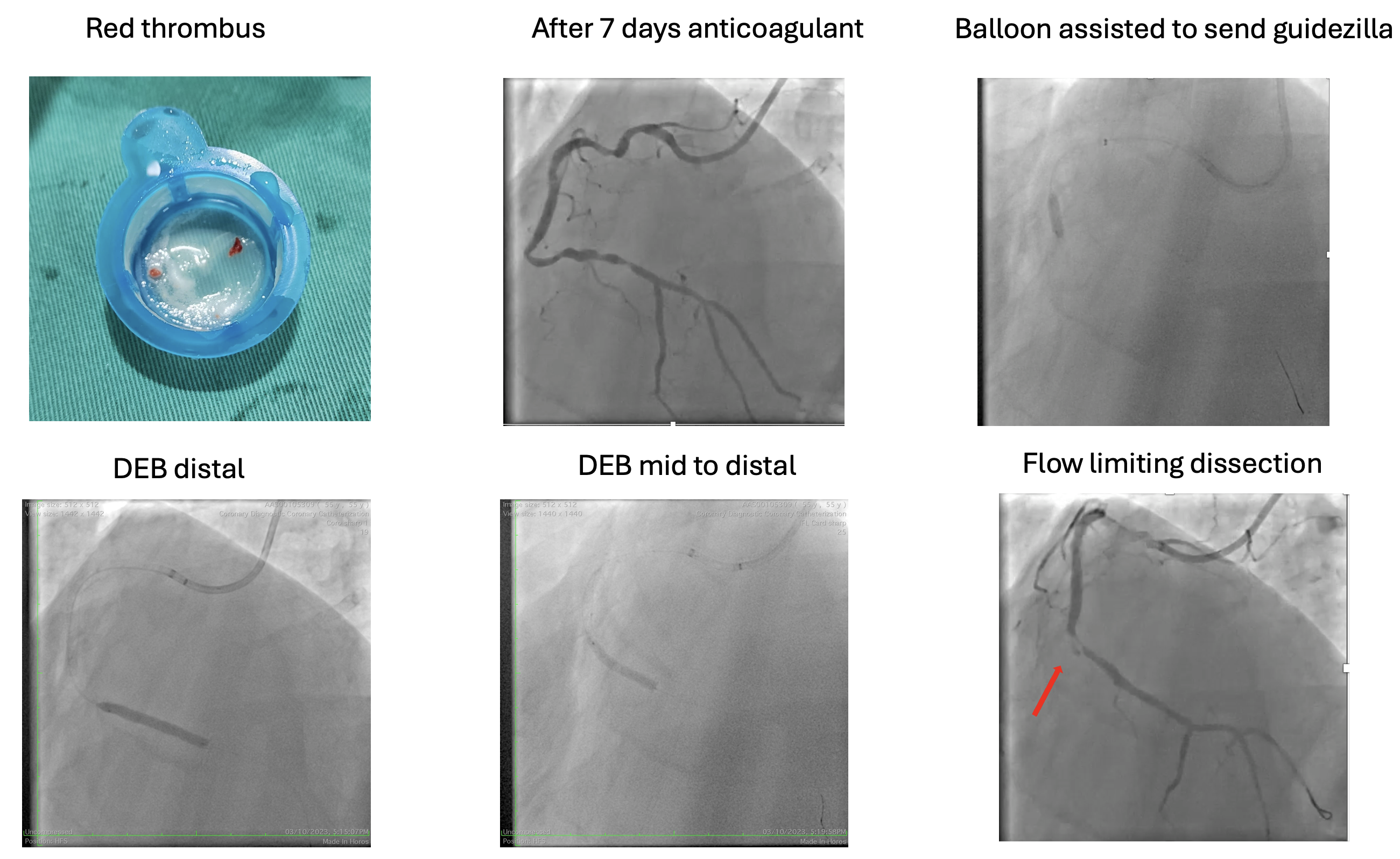
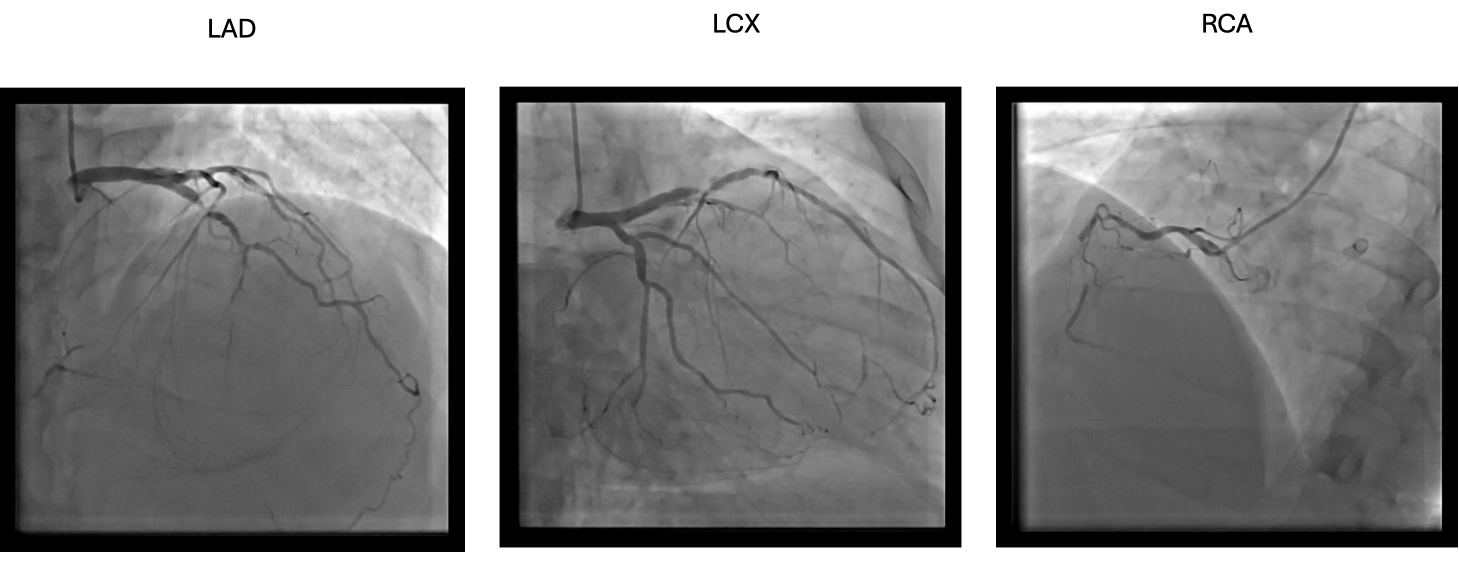
55 year old man with underlying severe plaque psoriasis on s/c ustekinumab had an anterior myocardial infarction in 2012 while travelling. He was not offered thromblysis and had only underwent angiogram 2 weeks later after returning and LAD was stented.  In 2023, he had NSTEMI and during that time, his ejection fraction had worsen to 30% and cardiac MRI showed non viable LAD. Repeated angiogram showed ISR 99% of LAD, recessive LCX and CTO dominant RCA. 

Relevant Test Results Prior to Catheterization

Cardiac MRI : Ischemic dilated cardiomyopathy with moderate LV systolic dysfunction ( LVEF 32% ) and normal RV systolic dysfunction ( RVEF 58% ). Minimal viability seen within mid to apical anterior, mid to apical anterior-septum and all apical segments ( Non viable mid LAD territory ) . All other segments are viable

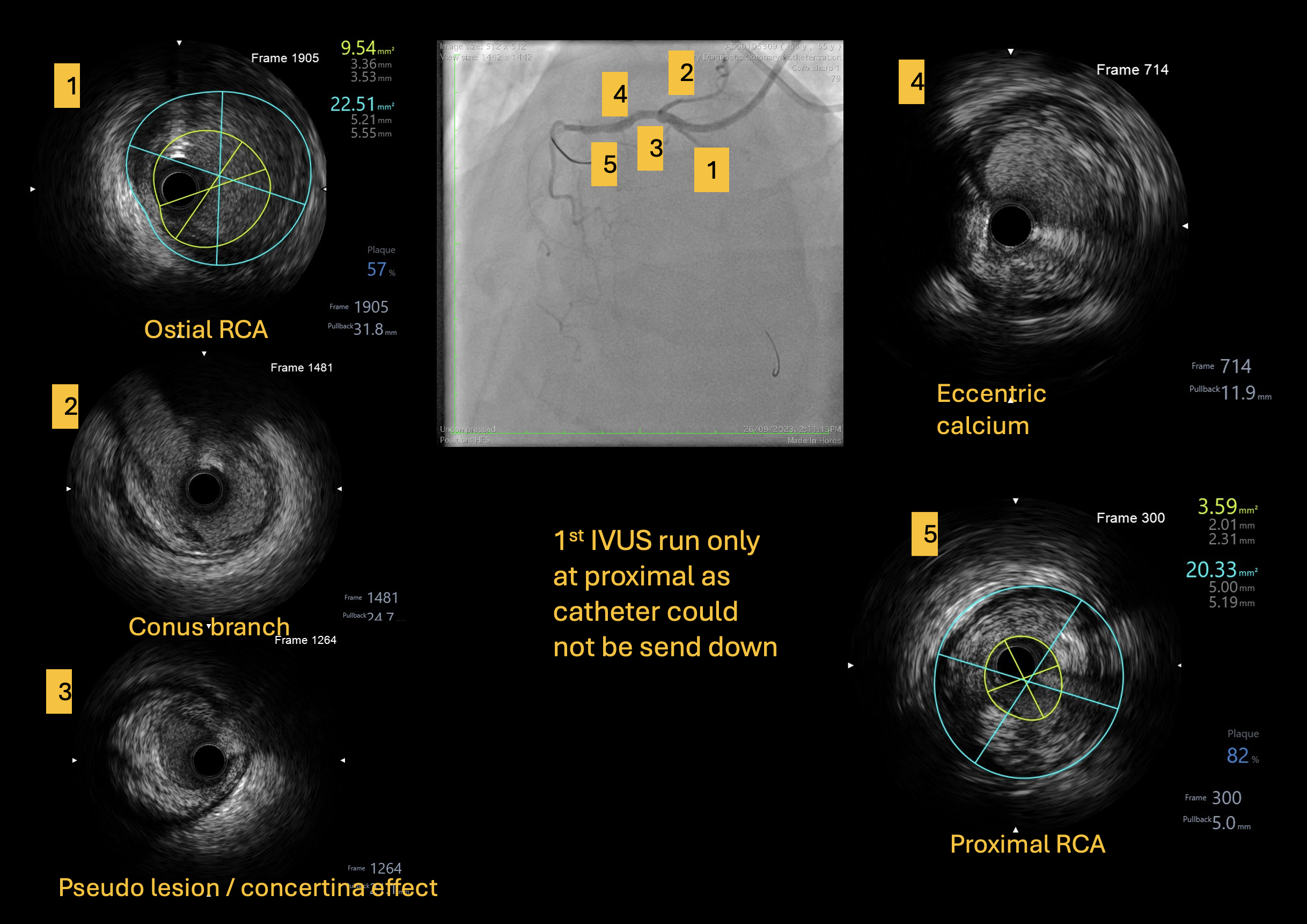
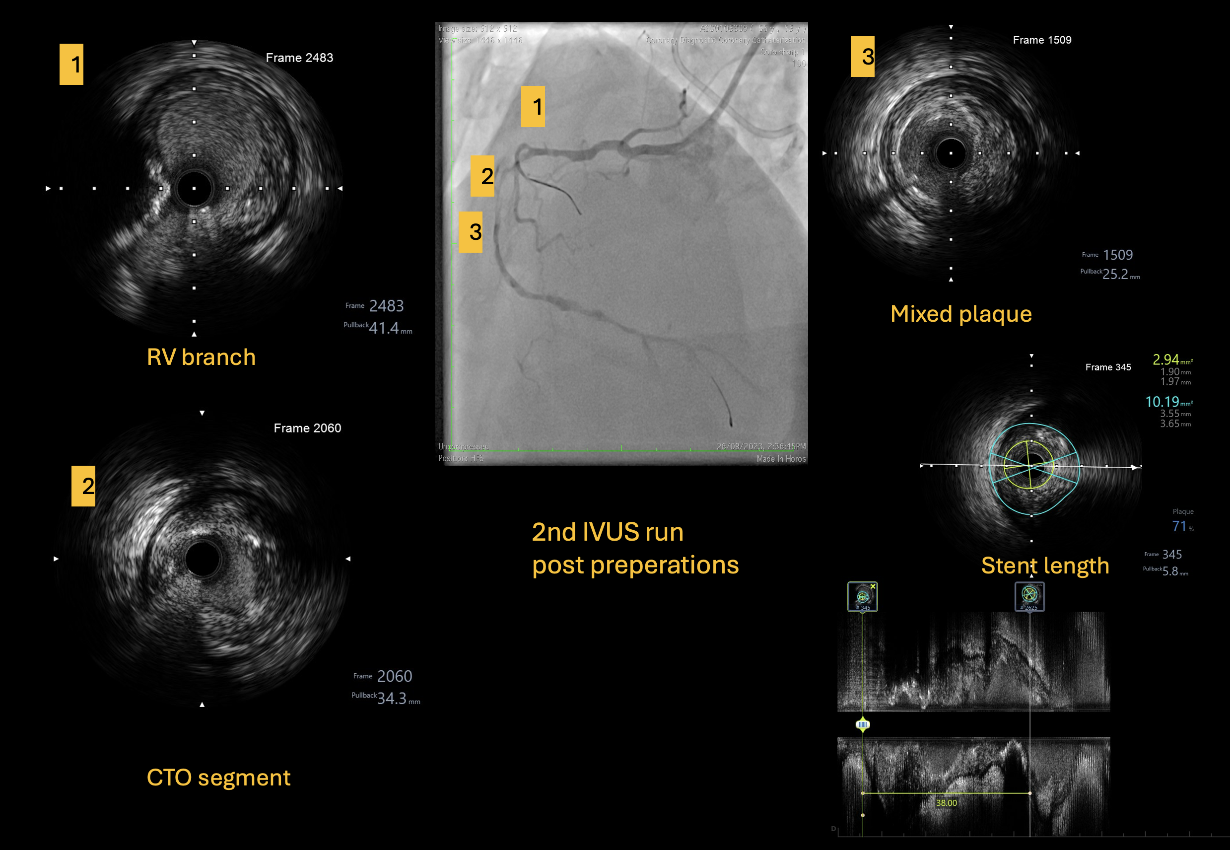
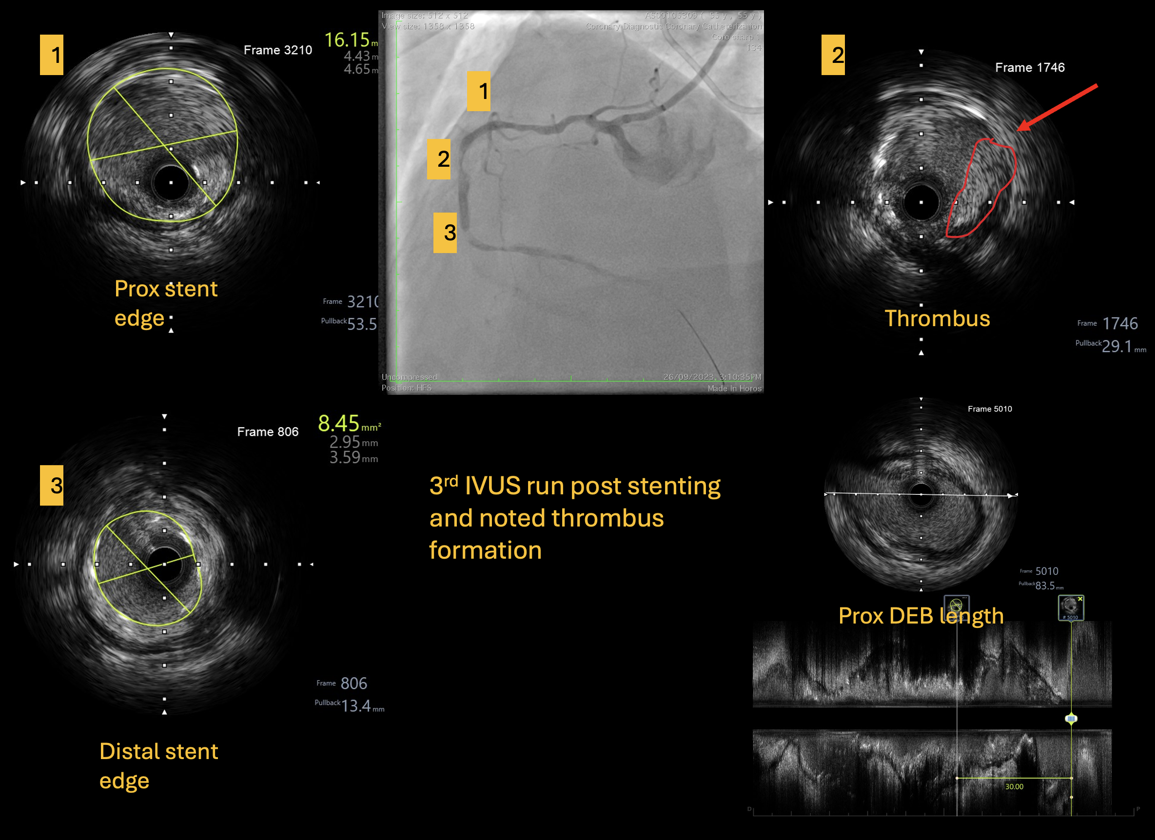
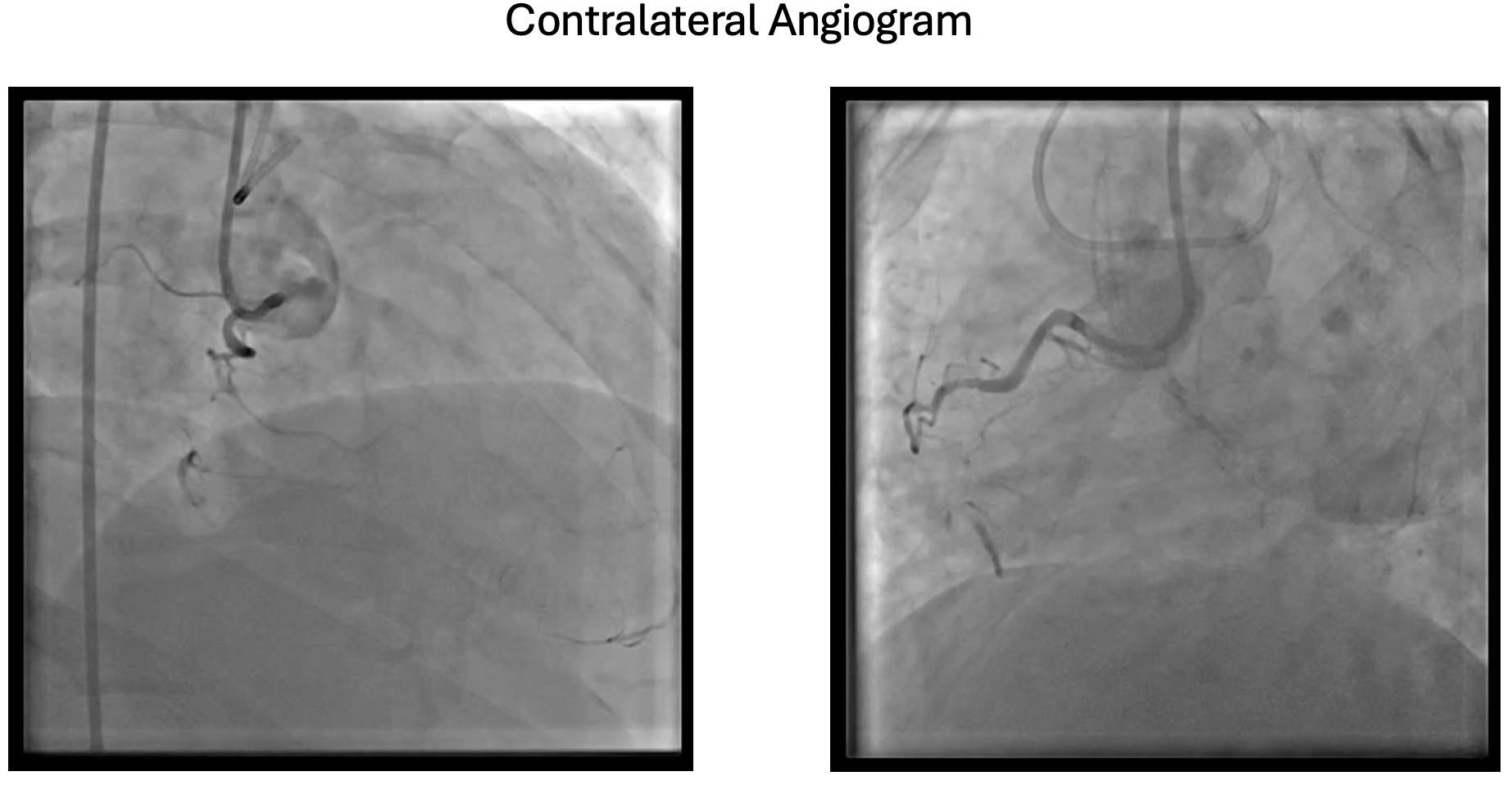
Relevant Catheterization Findings

His angiogram showed proximal LAD stent ISR 99%, mid LAD stent ISR 95%, LCX smooth and large dominant mid RCA chronic total occlusion with bridging collaterals. The J-CTO was 4 and the 1st attempt to intervene the CTO RCA by another operator failed.  Using IVUS and also branches of RCA, we determined our stent and DEB size and length. From the IVUS also noted pseudo lesion or concertina effect caused by the tortuous vessels.


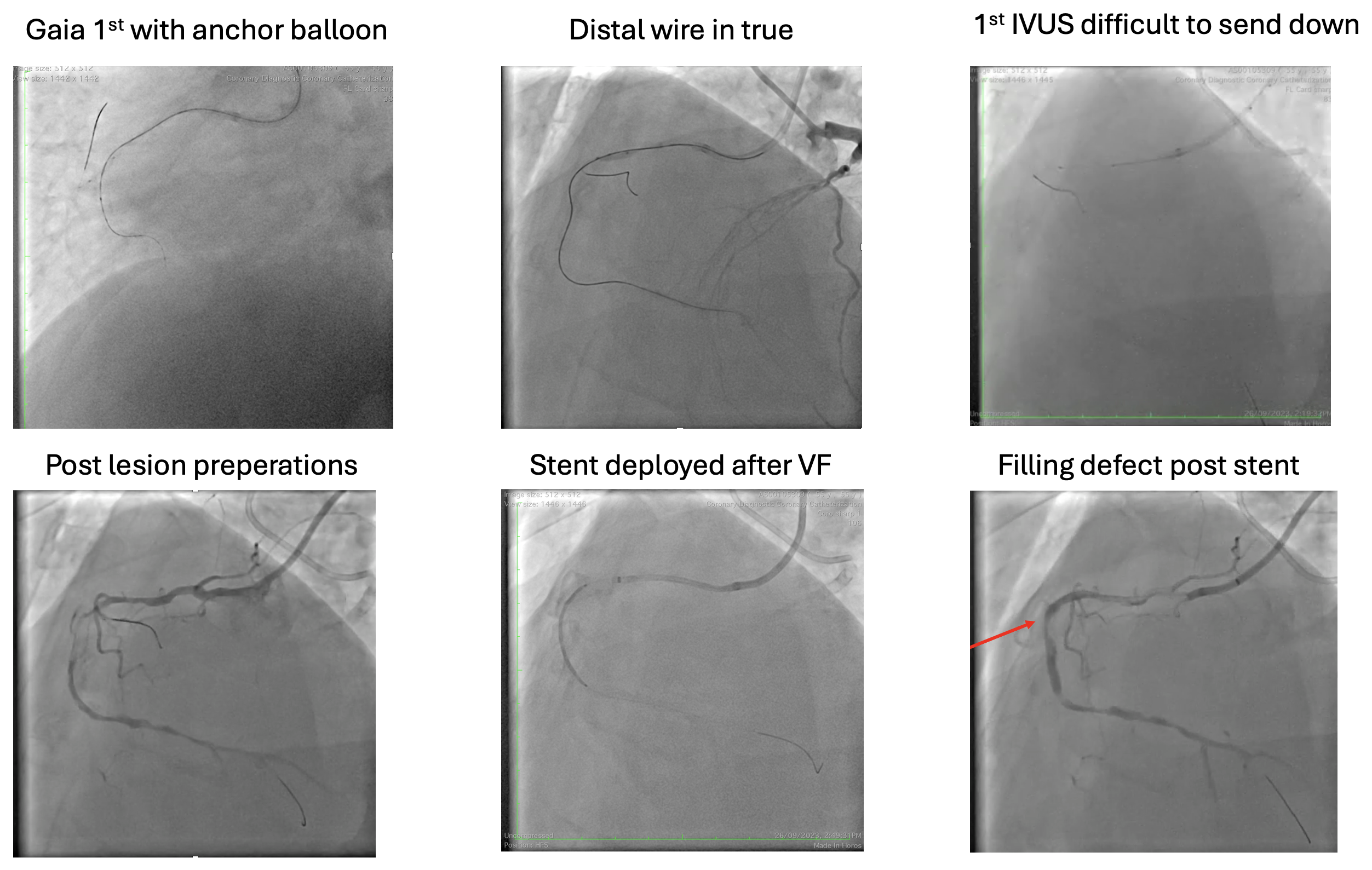
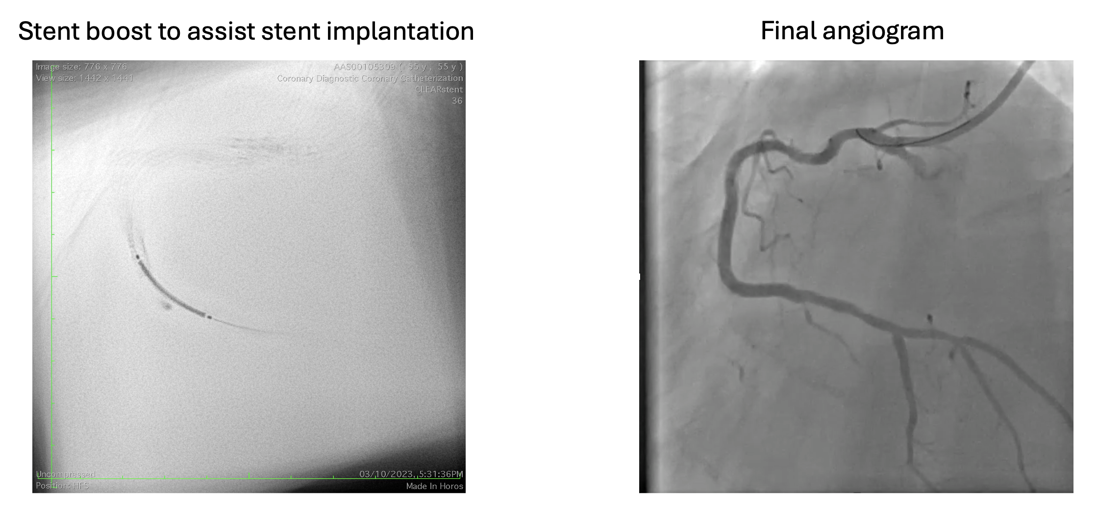
Interventional Management

Procedural Step

SAL used as guide for RCA and JL diagnostic for contralateral injections. Failed to cross with Fielder XTR in Finecross but successfully cross with Gaia 1st with an anchor balloon support due to tortousity. Unable to pass down Finecross thus lesion was prepared and wire was exchanged successfully. IVUS  was able to only advance till mRCA and noted diffuse disease from mid to proximal RCA. Further lesion prepared with 2.0 and 3.0 balloon. Able to send IVUS down further and noted attenuated plaque and vessel size of 4.5mm. In view of tortuosity, Guidezilla used to send a 3.5x38 stent. While advancing Guidezilla, there was pressure damping and patient developed chest pain and VF requiring cardioversion. Atropine and nitrates were given and stent was immediately deployed. With Guidezilla, stent post dilate with a 4.0 balloon. Noted filling defect at proximal stent and suspected thrombus. ACT remained more than 300 at all times. IC tirofiban was given and IVUS confirmed thrombus. Aspirations done showed red thrombus and IC streptokinase given. Decided to stop for proper anticoagulation. Relook coros after a week showed resolution of thrombus. pRCA, mRCA and dRCA prepared with a 3.5 scoring and 2 3.5x30 DCBs were deployed using Guidezilla with balloon assisted technique.  Another 3.5x25 DCB overlapped with distal stent segment. Wire removed and noted slow flow. Noted dissection at the distal RCA. Immediately rewire and stent with a 3.5x22 stent and post dilate with a 3.5x10 balloon.


Case Summary

Patient well and his EF had improved from 30% to 40% and now symptoms free. During the procedure, we wondered why patient had thrombosis despite ACTs were adequate. We found out that s/c ustekinumib, the biologics used for his psoriasis had been reported to have increased incidence of thrombosis. Other learning points in this case are tortuous vessel is the bane of intervention, even more so in CTOi. Next in tortuous vessels, wires, IVUS and Guidezilla can cause pseudo lesion, also known as concertina effect. DCB can be a good option in tortuous vessels if proper preparations is done and always be prepared with managing flow limiting dissection.