
Lots of interesting abstracts and cases were submitted for TCTAP 2022. Below are the accepted ones after a thorough review by our official reviewers. Don’t miss the opportunity to expand your knowledge and interact with authors as well as virtual participants by sharing your opinion in the comment section!
TCTAP A-057
Prone Contrast Enhancement Computed Tomography Angiography Protocol to Diagnose Concealed Endoleak and Aortic Sac Hygroma
By Sherif Sultan, Yogesh Acharya, Hesham Elzomor, Juan Parodi, Niamh Hynes, Osama Soliman
Presenter
Sherif Sultan
Authors
Sherif Sultan1, Yogesh Acharya1, Hesham Elzomor2, Juan Parodi3, Niamh Hynes2, Osama Soliman2
Affiliation
Western Vascular Institute, University Hospital Galway, Ireland1, National University of Ireland, Galway, Ireland2, University of Buen, Argentina3
View Study Report
TCTAP A-057
Imaging: Non-Invasive
Prone Contrast Enhancement Computed Tomography Angiography Protocol to Diagnose Concealed Endoleak and Aortic Sac Hygroma
Sherif Sultan1, Yogesh Acharya1, Hesham Elzomor2, Juan Parodi3, Niamh Hynes2, Osama Soliman2
Western Vascular Institute, University Hospital Galway, Ireland1, National University of Ireland, Galway, Ireland2, University of Buen, Argentina3
Background
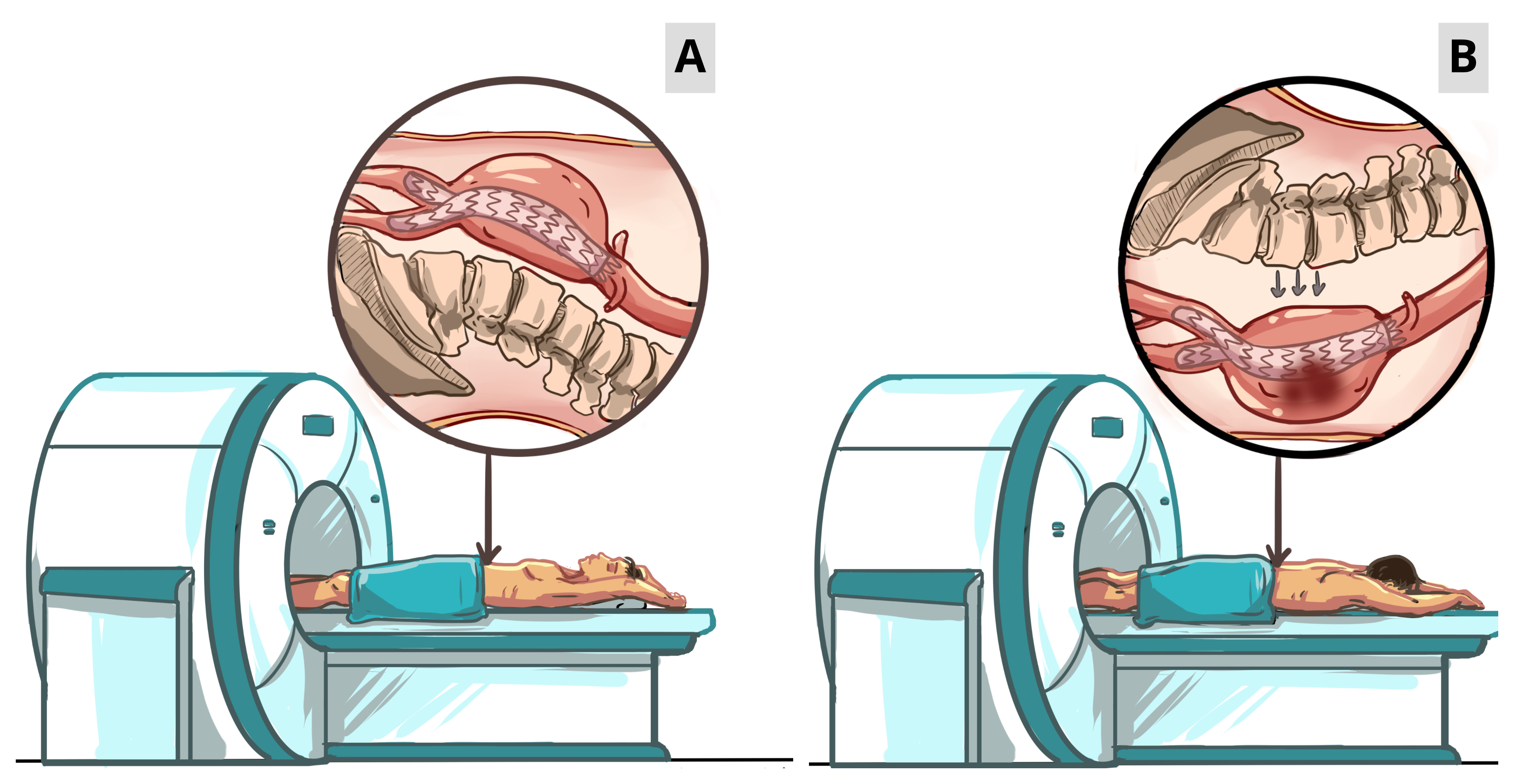
Standard 4-phase supine computed tomography angiography (CTA) scans are unable to detect smaller fabric tears (< 1 - 2 mm) following EVAR, which are erroneously interpreted as type IV or V endoleak (EL). Here, we propose a novel diagnostic technique, 'Prone contrASt enHanced computed tomography Angiography' (PASHA)' that uses time-resolved four-phase CTA protocol to detect and classify concealed ELs.
Methods
We employed PASHA protocol in 46 patients after continuous sac expansion following primary EVAR where the standard 4-phase CTA could not identify the EL source (Figure 1A). PASHA protocol uses a prone position during standard 4-phase CTA, which displaces the aorta anteriorly by approximately 5 mm, shortening the anterior cortex distance and allowing fluid transudation due to gravity with maximal EL enhancements (Figure 1B).
Results
The average number of CT scans performed before diagnosing EL with PASHA protocol were 3.4 ± 1.5 (range: 1 - 6). PASHA protocol demonstrated EL in all 46 cases that the standard supine 4-phase CTA could not identify. PASHA protocol effectively enhanced the degree of contrast infiltration into the aortic sac in all the patients.
Conclusion
PASHA is a multi-phase positional CTA technique that enhances the degree of contrast infiltration into the aortic sac when micro-leaks are present, increasing the EL detection rate following EVAR and strategically mapping successful future intervention.