
Lots of interesting abstracts and cases were submitted for TCTAP 2022. Below are the accepted ones after a thorough review by our official reviewers. Don’t miss the opportunity to expand your knowledge and interact with authors as well as virtual participants by sharing your opinion in the comment section!
TCTAP A-053
Multilayer Convolutional Neural Network Computer Vision is Effective in Automatic OCT Image Processing.
By Irina Alikovna Mustafina, Vladimir Ishmetov, Naufal Zagidullin, Igor V. Buzaev
Presenter
Irina Alikovna Mustafina
Authors
Irina Alikovna Mustafina1, Vladimir Ishmetov1, Naufal Zagidullin1, Igor V. Buzaev1
Affiliation
Bashkir State Medical University, Russian Federation1
View Study Report
TCTAP A-053
Imaging: Intravascular
Multilayer Convolutional Neural Network Computer Vision is Effective in Automatic OCT Image Processing.
Irina Alikovna Mustafina1, Vladimir Ishmetov1, Naufal Zagidullin1, Igor V. Buzaev1
Bashkir State Medical University, Russian Federation1
Background
The problem of automatic standardized interpretation OCT images is important due to the need for fast OCT-analysis during the procedure, as well as the operator's different experience in interpreting OCT images. Automatic analysis of the components of the atherosclerotic plaque will help in quick decision-making during operations on coronary vessels. The aim of the study was to evaluate the quality and efficiency of the automatic OCT image processing model by creating and training a neural network.
Methods
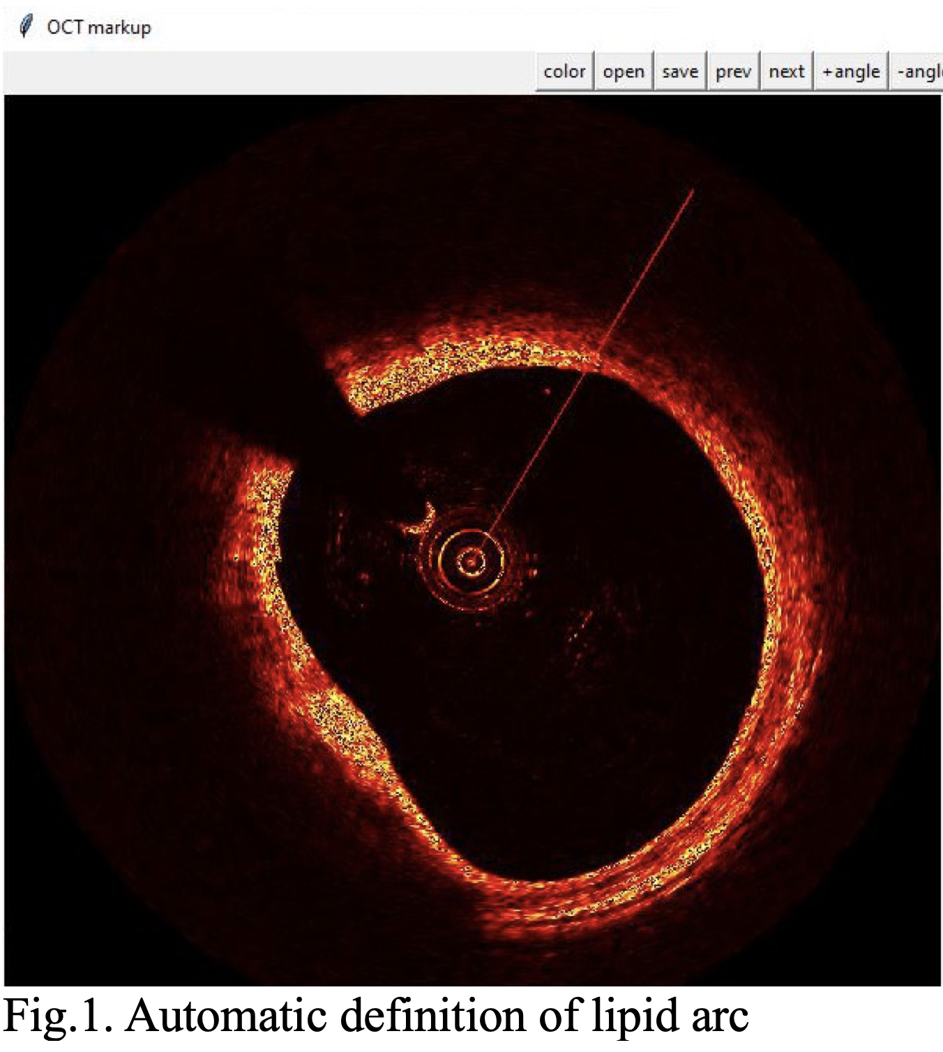
OCT images of patients with acute coronary syndrome and stable coronary artery disease were used for the neural network training matrix. The Python programming language was chosen to implement the software. To work with neural networks, the PyTorch library was used, developed and maintained by Facebook. Images were preprocessed using the OpenCV library. The GUI of the application was written using the Tkinter module from the Python standard library. To train neural network models, a total of 16 different types of pathologies were considered. To train classifiers, the expert marked up 534 images, 289 of which contained pathology. For training and validation of the binary classifier to detect the presence of pathology, the sample was divided into two parts: 426 images in the training sample (193 pathology / 233 norm) and 108 images for validation (56 pathology / 52 norm). The following samples were prepared for the lipid plaque classifier: 233 images in the training sample (157 lipid plaque / 76 other pathologies) and 56 images for control (40 lipid plaque / 16 other pathologies). The following samples were prepared for the fibrous plaque classifier: 233 images in the training sample (73 fibrous plaque / 160 other pathologies) and 56 images for control (16 fibrous plaque / 40 other pathologies) (Fig.1).
Results
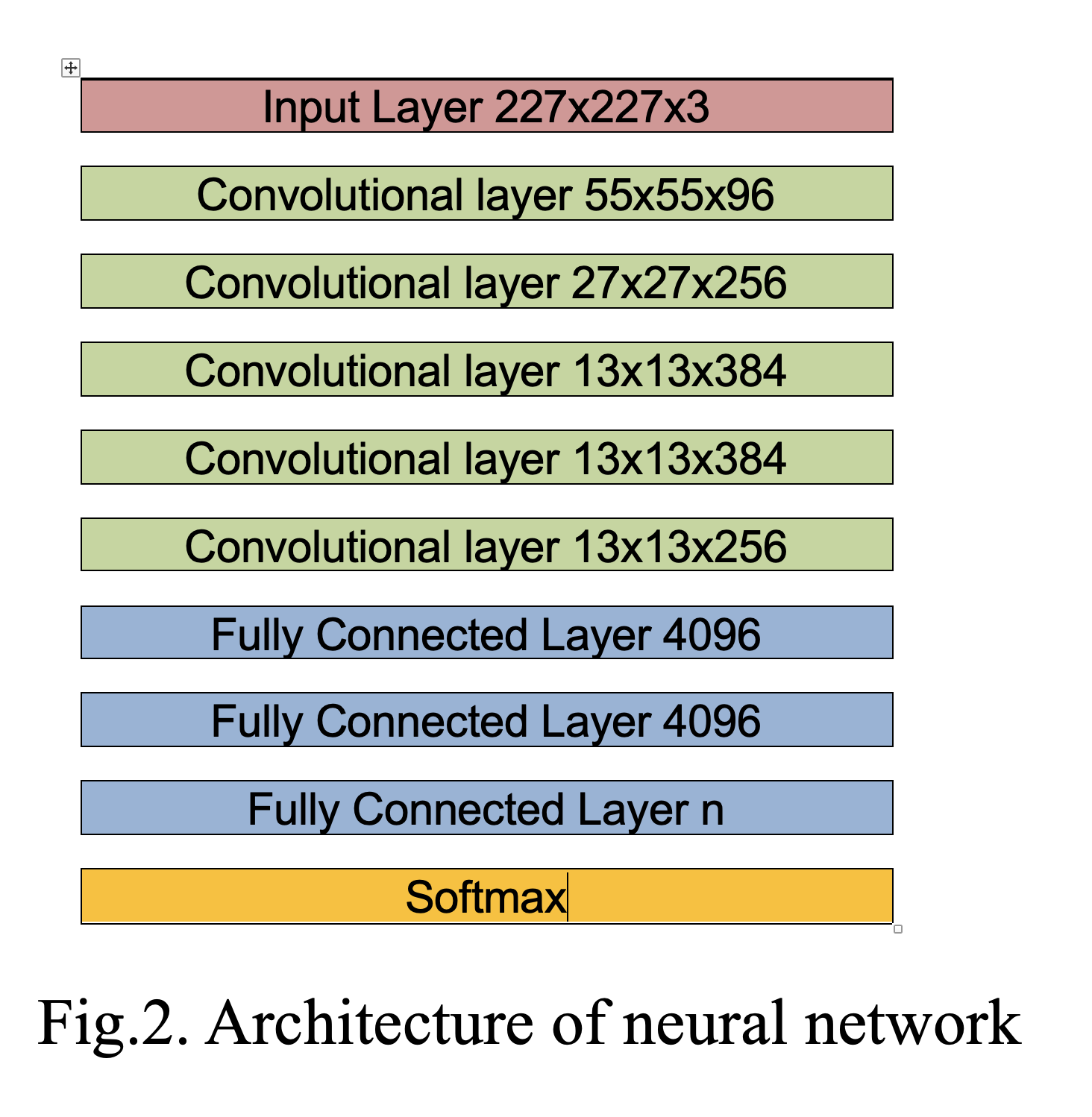
Three models were trained to solve the tasks set: 1) binary classifier for predicting the presence of pathologies; 2) binary classifier for predicting whether there is a fibrous plaque on the frame with pathology; 3) binary classifier for predicting whether there is a lipid plaque on the frame with pathology. Two common convolutional neural networks were considered as classifiers: AlexNet and ResNet-18 (Fig.2). Models pre-trained on the ImageNet dataset were taken and, with the help of the TransferLearning method, were retrained to solve the classification tasks. The training took place over 5 epochs. To detect coronary artery pathology, the accuracy was 0.97-1.0, to determine the presence of lipid plaque — 0.87 -0.92, to detect the presence of fibrous plaque 0.89-0.92.
Conclusion
A comprehensive software has been developed that allows separating a normal coronary artery from pathology, recognizing the type of atherosclerotic plaque, as well as marking the content of these components. Machine learning algorithms are capable of differentiating the lipid and fibrous components of atherosclerotic plaque with sufficiently high accuracy.