
Lots of interesting abstracts and cases were submitted for TCTAP 2022. Below are the accepted ones after a thorough review by our official reviewers. Don’t miss the opportunity to expand your knowledge and interact with authors as well as virtual participants by sharing your opinion in the comment section!
TCTAP A-063
Does Carotid Stenosis Lead to Caput Mandibulae Changes?
By Nataliya Makusheva, Sergey Chuykin, Vladimir Plechev, Luiza Temirova, Igor V. Buzaev, Pavel Malyshev
Presenter
Nataliya Makusheva
Authors
Nataliya Makusheva1, Sergey Chuykin1, Vladimir Plechev1, Luiza Temirova2, Igor V. Buzaev1, Pavel Malyshev2
Affiliation
Bashkir State Medical University, Russian Federation1, Republican Cardiology Center, Russian Federation2
View Study Report
TCTAP A-063
Others (Unclassified)
Does Carotid Stenosis Lead to Caput Mandibulae Changes?
Nataliya Makusheva1, Sergey Chuykin1, Vladimir Plechev1, Luiza Temirova2, Igor V. Buzaev1, Pavel Malyshev2
Bashkir State Medical University, Russian Federation1, Republican Cardiology Center, Russian Federation2
Background
There search was done with the aim to determine pathological changes in caput mandibulae in patients with stenosis of the external, common or bifurcationzone of carotids. The study included 46 female and male patients aged 50 to 65 years. The patients were divided into 2 groups - 30 and 16 patients with stenosis of the carotids, and without stenosis of carotids, but with atherosclerosis respectively. We studied angiograms to determine the presence and degree of stenosis of thecarotid arteries. We used the Hounsfield units for estimation of bone density by computed tomography, for densitometry of the caput mandibulae in patients ofboth groups. The research revealed: In the first group (with stenosis of the carotid arteries), the mean value of Hounsfield units on the right was 382.6 ±22.7, on the right - 388.7 ± 19.2. In the second group (without stenosis), themean value of Hounsfield units on the right was 489.7 ± 18.1; on left - 490.8 ±17.2. There is a high correlation between carotid narrowing and caput mandibulae density (p <0.05). Thas, the ischemic changes, a consequence of stenotic lesions of the carotids, lead to pathological changes in the bone tissue of the caput mandibulae.
Methods
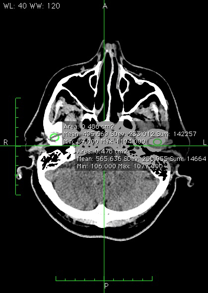
The study included 46 female and male patientsaged 66,1 ± 1,67 years. The first group included 30 patients with atherosclerotic stenosis of the external, common carotids or bifurcation zone (group 1 – with stenosis). The study included patients with hemodynamically significant stenosis - on average 61.1 ±2.94% on the right and 66.3 ± 2.85% on the left. The minimum stenosis value on the left is 30%; the maximum stenosis value is 100%.The second group included 16 patients 66,3 ±1,69 age, who had atherosclerosis, but does not have significant stenosis of the main arteries goes to the organs of the oral cavity (group 2 – without stenosis). To study the degree of stenosis of the carotids, we investigated angiography and ultrasound duplex scanning (Republican Cardiology Centre, Ufa).To determine differences, we took patients with significant stenosis on one or two sides. However, there were patients with a significant path with one and insignificant on the other.We examined computed tomograms of patients in both groups. Hounsfield units were measuredat caput mandibulae on the right and left sides (Fig. 1).
Results
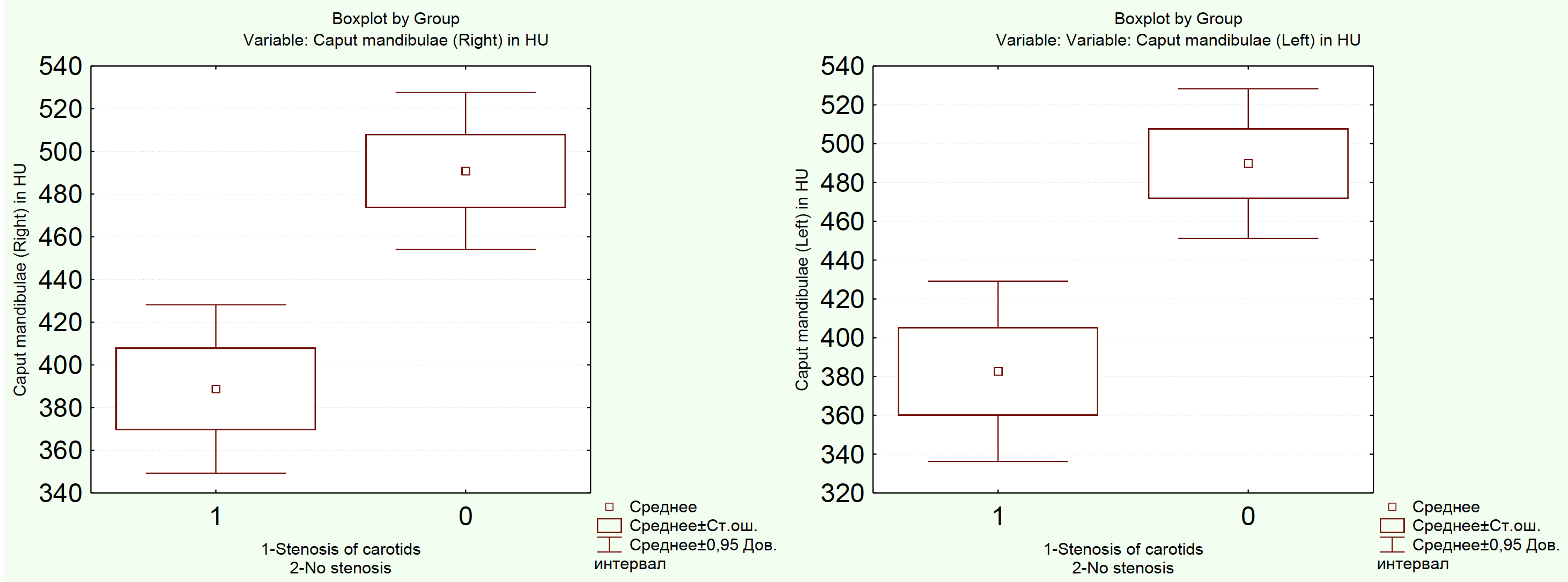
In the first group (with stenosis of the carotid arteries), the meanvalue of Hounsfield units on the right was 382.6 ± 22.7, on the left - 388.7 ±19.2. In the second group (without stenosis), the mean value of Hounsfield units on the right was 489.7 ± 18.1; on left - 490.8 ± 17.2. We noted a decreasein Hounsfield units in patients of the first group with stenosis compared with patients of the second group (Tab. 2).
When comparing the two groups, Boxplot by Mann-Whitney shows that there is a high correlation between carotid narrowing and caput mandibulae density (p <0.05) (Fig. 2).
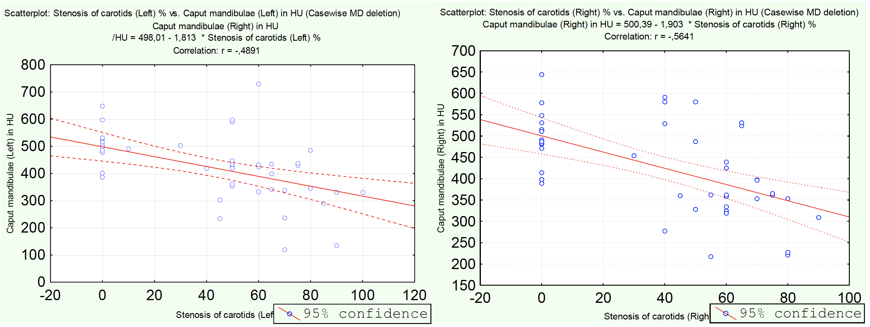
We made the scatterplot of the Hounsfield units in the caputmandibulae depending on the degree of narrowing of the carotids in the patients of the first group on the right and on the left. There is a pronounced correlation with the correlation coefficient r = 0.48 and r = 0.56, respectively (Fig. 3).
The obtained results of the correlation analysis indicate the dependence ofpathological changes in the caput mandibulae on the degree of atheroscleroticnarrowing of the external carotids, common carotids or bifurcation zone.
| M | CI -95% | CI +95% | Min | Max | σ | m | ||
| 1 group | Caput mandibulae Left | 382,67 | 336,22 | 429,12 | 120,0 | 730,0 | 124,39 | 22,71 |
| 1 group | Caput mandibulae Right | 388,73 | 349,28 | 428,18 | 217,0 | 592,0 | 106,65 | 19,29 |
| 2 group | Caput mandibulae Left | 489,75 | 451,17 | 528,33 | 386,0 | 648,0 | 72,41 | 18,10 |
| 2 group | Caput mandibulae Right | 490,81 | 454,01 | 527,61 | 389,0 | 644,0 | 69,06 | 17,27 |
Conclusion
Thus, when comparing groups of patients with atherosclerotic stenosis of the common or external carotid arteries, or bifurcation zone of carotids (group 1) and with outstenosis of carotids (group 2), we obtained results indicating statistically differences in the pathology of caput mandibulae in patients of 1 group.
Our data indicate that ischemic changes, a consequence of stenotic lesions of the carotids, lead to pathological changes in the bone tissue of the caput mandibulae.