
Lots of interesting abstracts and cases were submitted for TCTAP 2023. Below are the accepted ones after a thorough review by our official reviewers. Don’t miss the opportunity to expand your knowledge and interact with authors as well as virtual participants by sharing your opinion in the comment section!
TCTAP C-163
A Rare Finding in Pregnancy Related Acute Coronary Syndrome
By Muhammad Hanis Muhmad Hamidi, Huzairi Sani, Ahmad Bakhtiar Md Radzi, Khairul Shafiq Ibrahim, Johan Rizwal Ismail, Abdul Wahab Undok
Presenter
Muhammad Hanis Muhmad Hamidi
Authors
Muhammad Hanis Muhmad Hamidi1, Huzairi Sani1, Ahmad Bakhtiar Md Radzi1, Khairul Shafiq Ibrahim1, Johan Rizwal Ismail2, Abdul Wahab Undok3
Affiliation
Universiti Teknologi MARA (UiTM), Malaysia1, Prince Court Medical Centre, Malaysia2, Kpj Rawang Specialist Hospital, Malaysia3,
View Study Report
TCTAP C-163
IMAGING AND PHYSIOLOGIC LESION ASSESSMENT - Imaging: Intravascular
A Rare Finding in Pregnancy Related Acute Coronary Syndrome
Muhammad Hanis Muhmad Hamidi1, Huzairi Sani1, Ahmad Bakhtiar Md Radzi1, Khairul Shafiq Ibrahim1, Johan Rizwal Ismail2, Abdul Wahab Undok3
Universiti Teknologi MARA (UiTM), Malaysia1, Prince Court Medical Centre, Malaysia2, Kpj Rawang Specialist Hospital, Malaysia3,
Clinical Information
Patient initials or Identifier Number
NM
Relevant Clinical History and Physical Exam
A 42y.o lady who is 4 weeks post-partum presented with sudden onset typical chest pain at rest not resolving with sublingual GTN. She is known diabetic with very high-risk pregnancy due to pre-existing IHD under a different center follow-up and has been uneventful to date. She had history of inferior MI 2 years ago where she underwent percutaneous coronary intervention with DOAC+DAPT for 6months and DOAC+SAPT for 6months. She is only on aspirin and bisoprolol throughout pregnancy.
Relevant Test Results Prior to Catheterization
We managed to trace her previous angiogram done 2 years ago (24/12/20) which showed thrombus in RCA with diffusely ectatic and atherosclerotic RCA,LCx and LAD disease. A conservative approach with POBA to RCA and anticoagulants were used back then to manage her coronary artery disease. Her ECHO at 36weeks gestation showed LVEF 50% with no regional wall motion abnormalities.
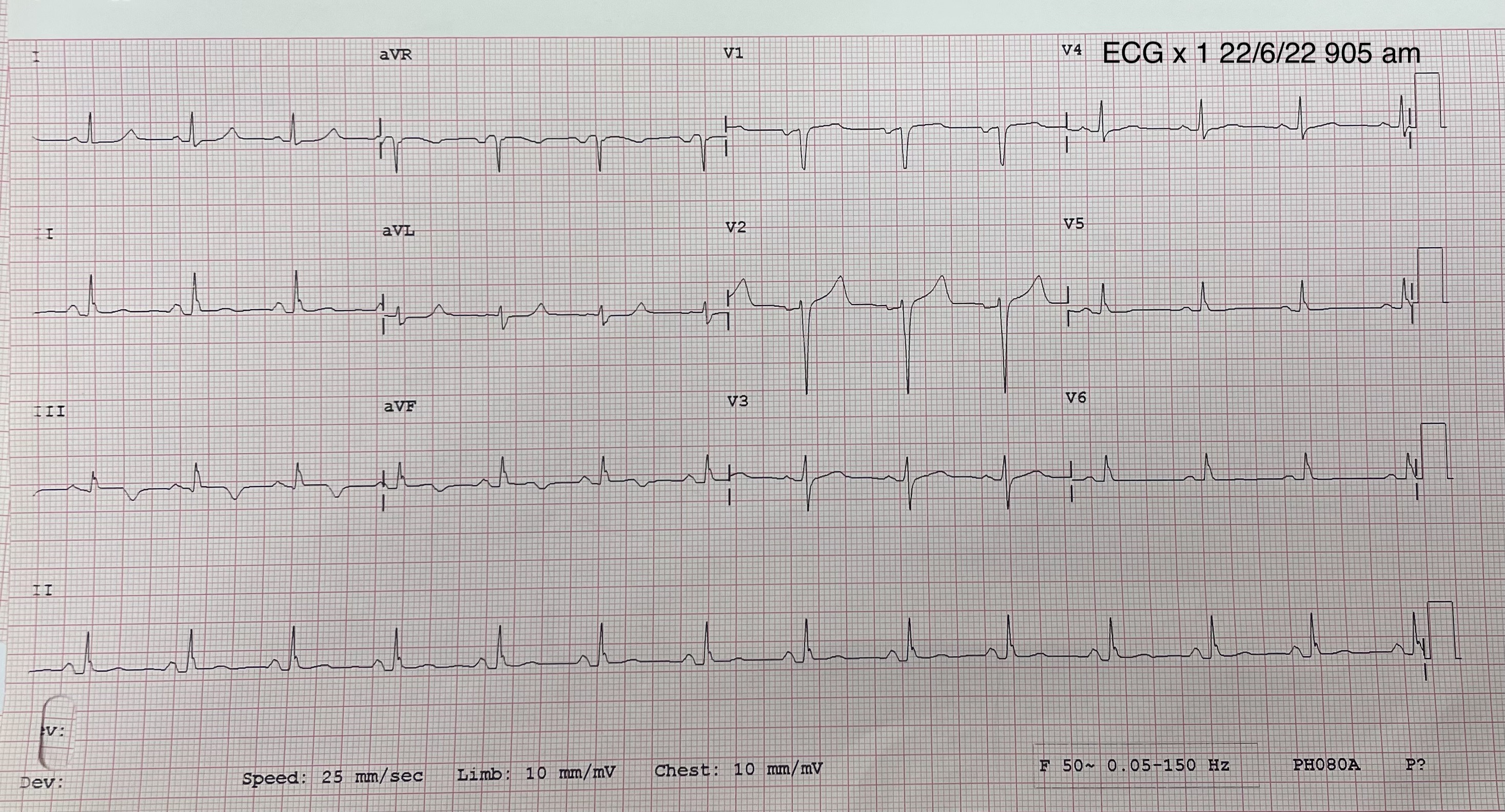
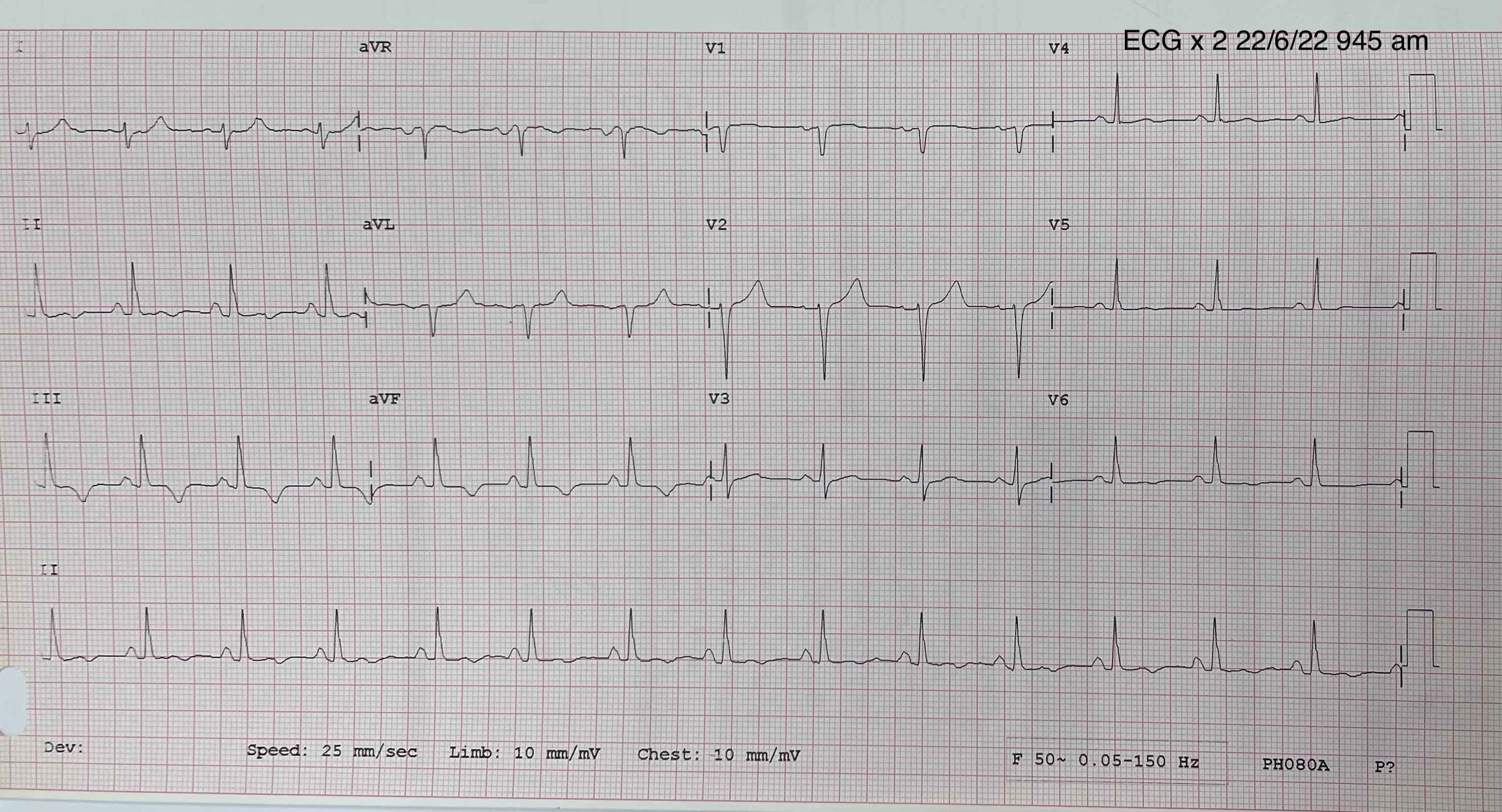
22/6/22Serial ECG: Deepening T inv III,aVFTroponin I : 14300pg/mLECHO : LVEF 45% with hypokinesia basal to mid inferior wall
22/6/22Serial ECG: Deepening T inv III,aVFTroponin I : 14300pg/mLECHO : LVEF 45% with hypokinesia basal to mid inferior wall
Relevant Catheterization Findings
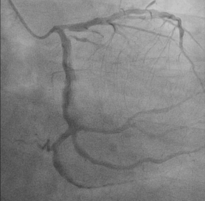
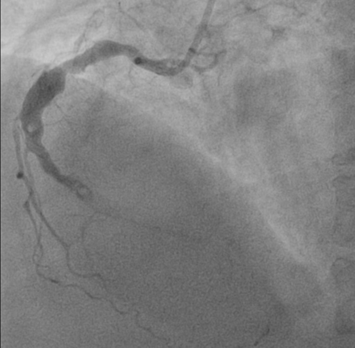
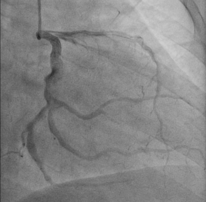
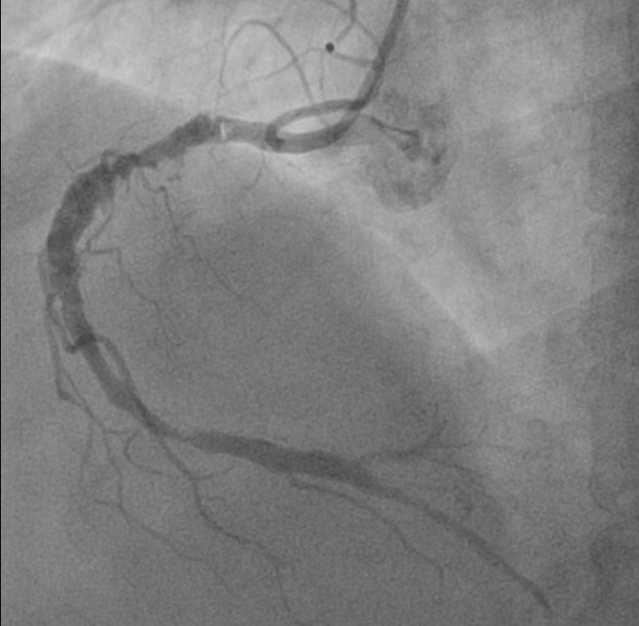
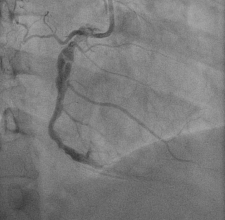
Urgent coronary angiogram were carried out in view of very high risk NSTEMI (persistent chest pain, dynamic ECG changes with very high Troponin). Coronary angiogram showed diffusely ectatic and atherosclerotic RCA,LCx and LAD disease. There is a woven-like appearance of proximal to mid RCA with intraluminal haziness and stenotic lesion at distal RCA.
Interventional Management
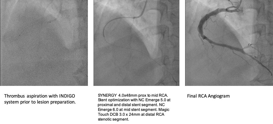
Procedural Step
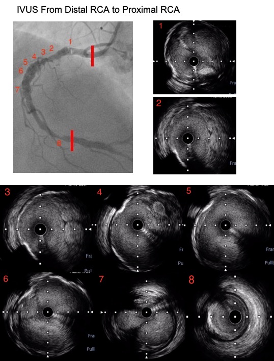
Intraluminal haziness from the angiogram is likely to be thrombus in this setting but spontaneous coronary artery dissection and ruptured plaque can be other highly likely possibilities. Thus, intravascular ultrasound (OptiCross HD, Boston Scientific) is used to ascertain the morphology and to determine the ectatic vessel size for PCI optimization.
Case Summary
Angiographically hazy coronary lesions are non-specific. Multiple linear filling defects are commonly described angiogram finding in coronary dissections but as shown in our case they can be the result of a chronic recanalized thrombus. Recanalized thrombus of coronary arteries is an under-recognized entity. Intravascular imaging conclusively establishes its diagnosis, which otherwise is often misdiagnosed as fresh thrombus, spontaneous coronary artery dissection, or severe calcification based on angiography alone.