E-Case

JACC

Lots of interesting abstracts and cases were submitted for TCTAP 2023. Below are the accepted ones after a thorough review by our official reviewers. Don¡¯t miss the opportunity to expand your knowledge and interact with authors as well as virtual participants by sharing your opinion in the comment section!

TCTAP C-005

An Evil From the Past

By Kai Soon Liew, Vijayendran Rajalingam, Yuen Hoong Phang, Pirevina Naidu Krishnan Naidu, Tamim Ansari Bin Jahubar Sathik, Izzatul Nadzirah Binti Ismail, Nur Asmalina Binti Azizan, Kantha Rao Narasamuloo, Saravanan Krishinan, Chee Tat Liew, Dharmaraj Karthikesan

Presenter

Liew Kai Soon

Authors

Kai Soon Liew1, Vijayendran Rajalingam2, Yuen Hoong Phang1, Pirevina Naidu Krishnan Naidu3, Tamim Ansari Bin Jahubar Sathik3, Izzatul Nadzirah Binti Ismail3, Nur Asmalina Binti Azizan3, Kantha Rao Narasamuloo1, Saravanan Krishinan4, Chee Tat Liew5, Dharmaraj Karthikesan3

Affiliation

Sultanah Bahiyah Hospital, Malaysia1, Sultan Idris Shah Serdang Hospital, Malaysia2, Hospital Sultanah Bahiyah, Malaysia3, Ministry of Health Malaysia, Malaysia4, Pantai Penang Hospital, Malaysia5,
View Study Report
TCTAP C-005
CORONARY - Acute Coronary Syndromes (STEMI, NSTE-ACS)

An Evil From the Past

Kai Soon Liew1, Vijayendran Rajalingam2, Yuen Hoong Phang1, Pirevina Naidu Krishnan Naidu3, Tamim Ansari Bin Jahubar Sathik3, Izzatul Nadzirah Binti Ismail3, Nur Asmalina Binti Azizan3, Kantha Rao Narasamuloo1, Saravanan Krishinan4, Chee Tat Liew5, Dharmaraj Karthikesan3

Sultanah Bahiyah Hospital, Malaysia1, Sultan Idris Shah Serdang Hospital, Malaysia2, Hospital Sultanah Bahiyah, Malaysia3, Ministry of Health Malaysia, Malaysia4, Pantai Penang Hospital, Malaysia5,

Clinical Information

Patient initials or Identifier Number

Mr X

Relevant Clinical History and Physical Exam

A 55 year old man with multiple risk factors presented to the district hospital with acute inferior posterior STEMI. He has a history of PCI to RCA in 2012 with drug eluting stents. Thrombolysis was given as per protocol and showed successful reperfusion on ECG. He developed ventricular fibrillation post thrombolysis and was resuscitated and intubated. ECG the next day showed reinfarction with cardiogenic shock and he was transferred to our cardiac centre for emergency revascularization.  


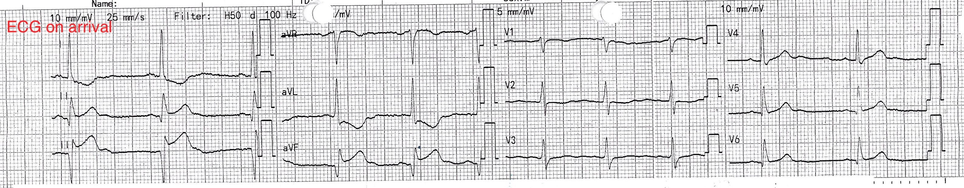
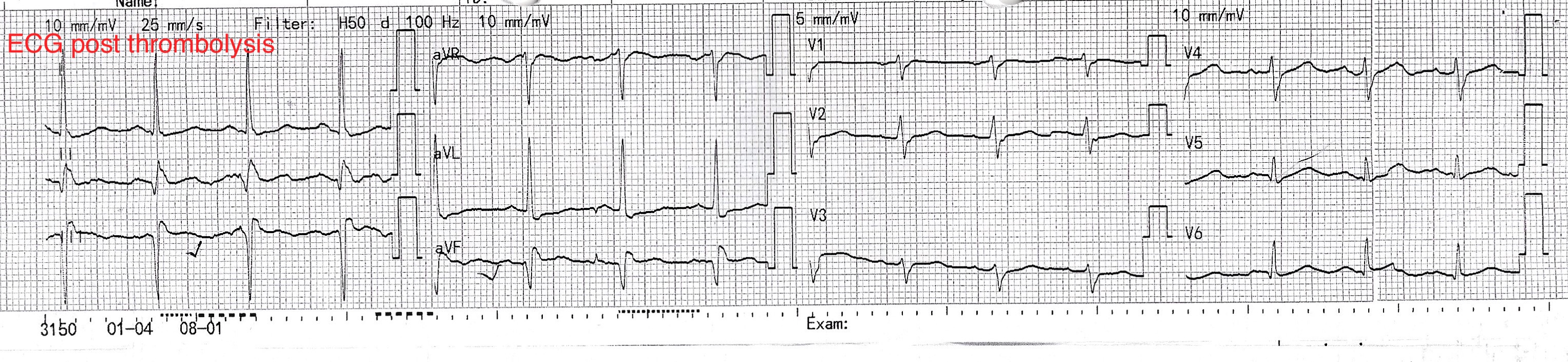
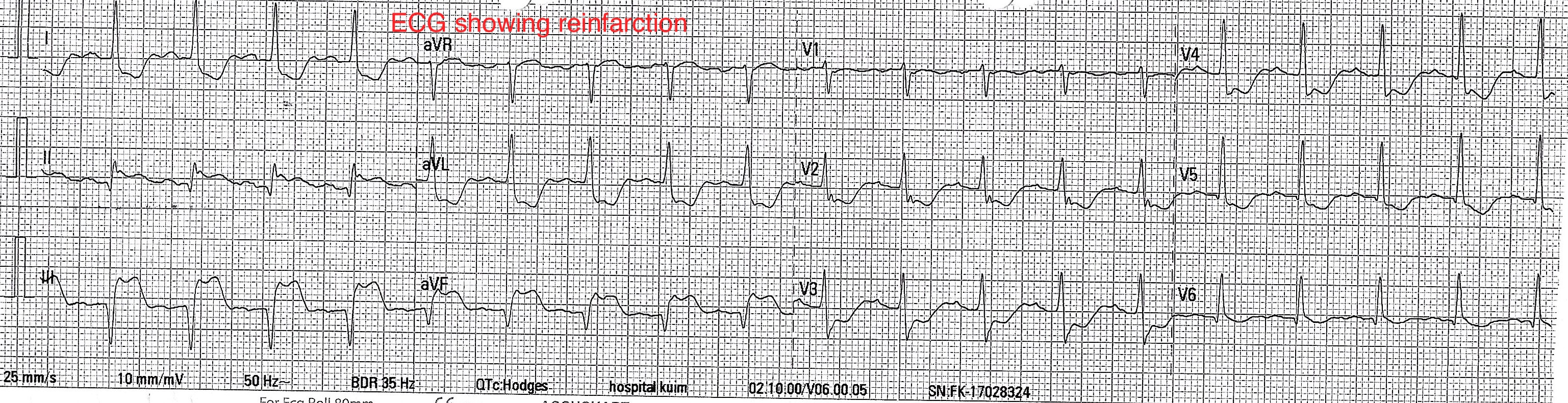
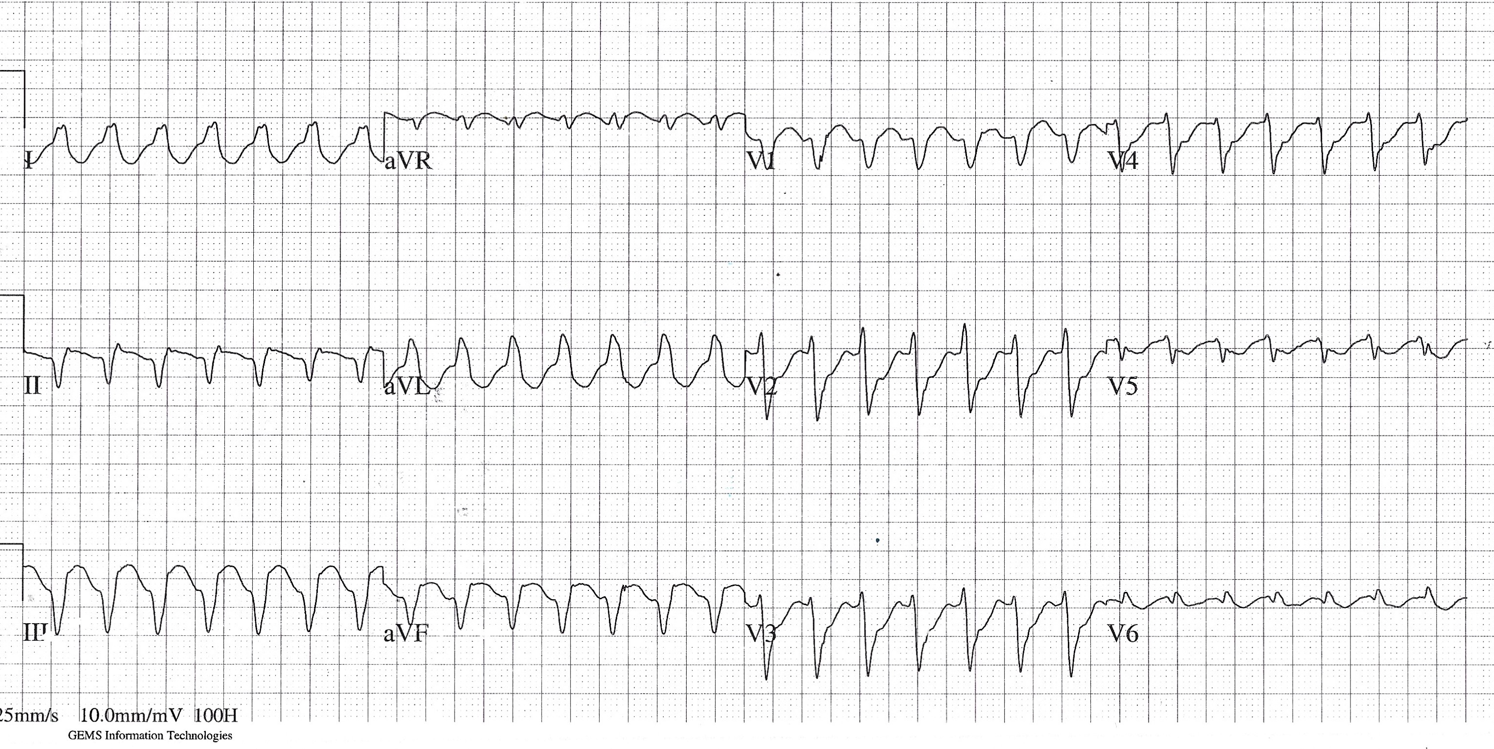
Relevant Test Results Prior to Catheterization

ECG on arrival showed persistent ST segment elevation over the inferior and posterior leads. Troponin was raised and eGFR was 39. He arrived to the catherization lab with pulseless electrical activity (PEA) and femoral access was obtained while CPR was in progress.  Runs of ventricular tachycardia was also observed, requiring multiple episodes of cardioversion during procedure. 

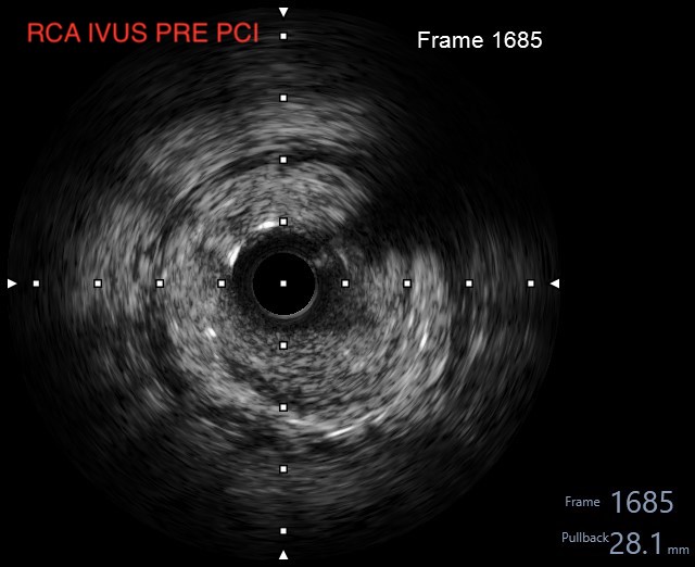
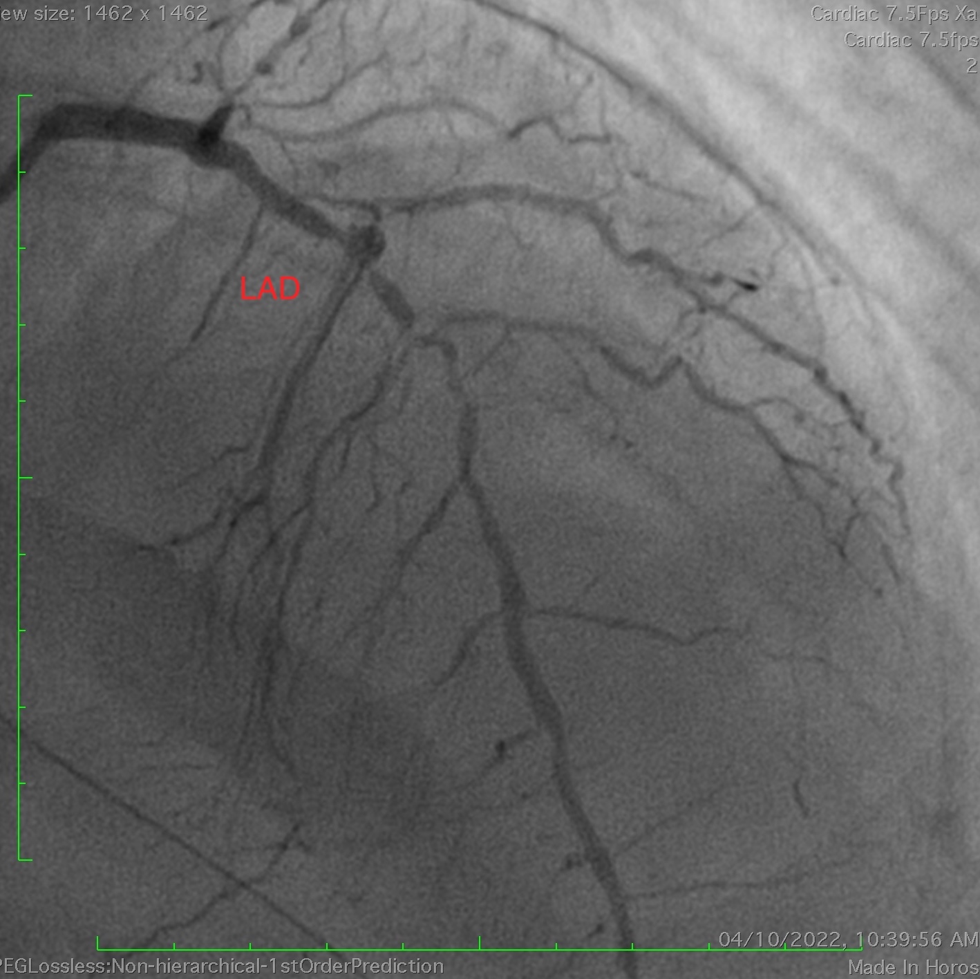
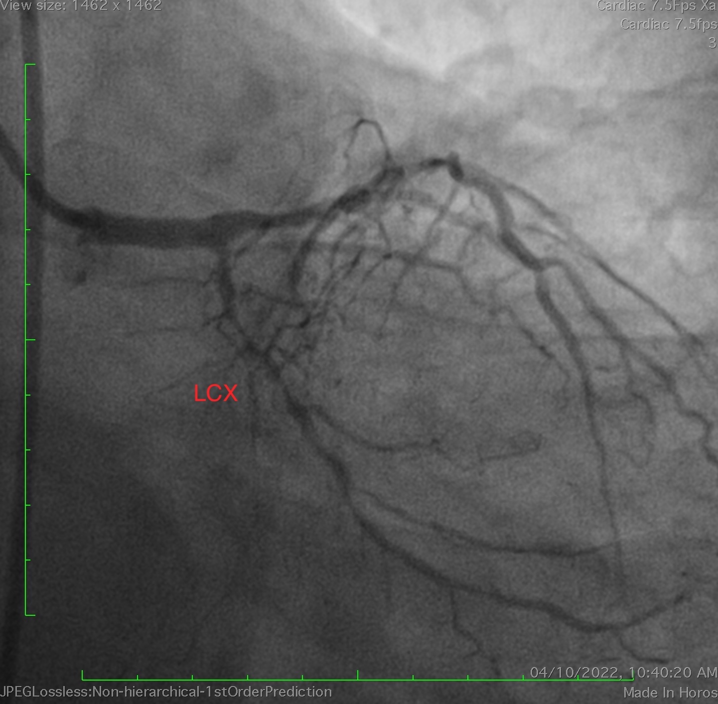
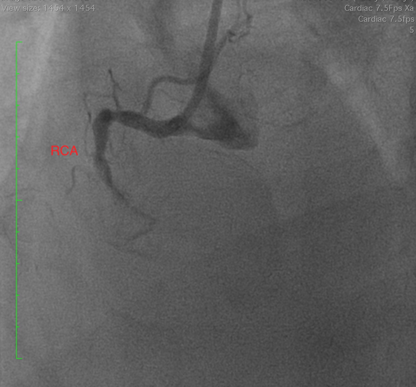
Relevant Catheterization Findings

Angiogram showed significant disease in mid left anterior descending artery (LAD) with diffuse disease in the left circumflex artery (LCX). Mid right coronary artery (RCA) was occluded with faint septal collaterals from LAD. RCA occlusion appears proximal to the previous stents on angiography. 


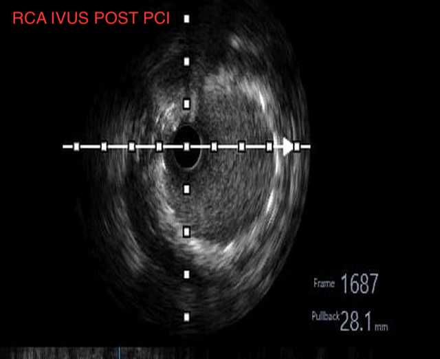
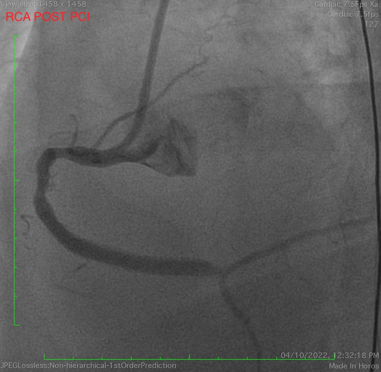
Interventional Management

Procedural Step

We proceeded with an emergency PCI to RCA with JR 4.0. We attempted with Sion Blue to RCA but our wire entered dissection plane. Using buddy wire technique, Sion black wire was wired into PLV. Lesion was predilated and using a dual lumen catheter, Sion blue wire was wired into PDA. IVUS assessment showed previous stents placed just before the bifurcation were undersized with heavy thrombus burden and in stent restenosis (ISR). Thrombus aspiration was done multiple times and TIMI 3 flow restored. ECG showed resolution of ST segment elevation. We decided to stop and relook later but patient developed multiple episodes of ventricular tachycardia right after removing the wires and angiography showed slow flow distally probably due to thrombus or plaque shift. Immediately rewired with dual lumen catheter support. RCA lesion distal to stent was prepared with Scoreflex 3.0x15 and Sapphire 3.5 x 15 non-compliant balloon was used to expand the previous undersized stents. IVUS assessment showed good lesion preparation and expansion of previous stents. A drug coated balloon (DCB) 3.0x15 was deployed at distal RCA to PLV. The previous stented segments were overlapped with a 4.0x48 drug eluting stent and post dilated accordingly. Final IVUS assessment showed excellent results. There was immediate resolution of ventricular tachycardia and ST segment elevation post PCI.


Case Summary

He was subsequently weaned off ventilator and inotropic support. His ejection fraction via echocardiography and renal function showed marked improvement and he was discharged well. He underwent stage PCI to LAD later on and relook to RCA stent remained well patent. Very late stent thrombosis can occur many years after DES implantation. Undersized stents are a major risk factor for late lumen loss. Coronary imaging using IVUS is an important tool to allow lesion assessment and post PCI optimization while minimizing contrast usage. In patients with impaired renal function, it is important to minimize contrast usage to prevent contrast nephropathy which ultimately leads to poorer prognosis